Sistema urinário: dos rins à uretra, quando a geometria decide o risco
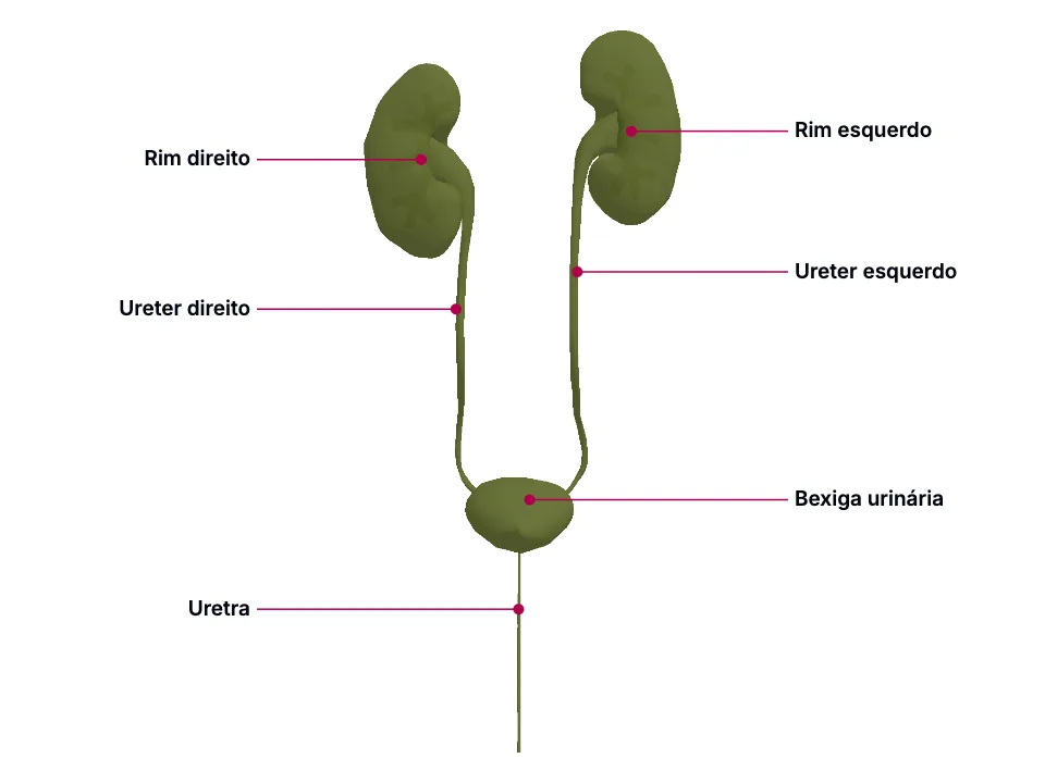
Do rim ao meato uretral, seis peças do trato urinário compartilham um padrão: um detalhe de FORMA - uma assimetria de trajeto venoso, um ponto de estreitamento verdadeiro, um ângulo de entrada, uma diferença de comprimento entre os sexos - decide um desfecho clínico real, da síndrome do quebra-nozes e do varicocele assimétrico à impactação de cálculo e ao refluxo vesicoureteral. Ureter e trígono vesical compartilham origem no ducto mesonéfrico, o que explica por que o ureter atravessa a parede da bexiga obliquamente, criando um mecanismo antirrefluxo sem esfíncter muscular dedicado. Por fim, a diferença de comprimento entre a uretra masculina (~20 cm, quatro segmentos) e a feminina, bem mais curta, é a explicação mecanística - não só estatística - da maior incidência de infecção urinária ascendente em mulheres.
Educational material written with AI support and checked by automated review, without a named medical reviewer. It does not replace the textbook or clinical guidance.
Essential
Os rins são órgãos retroperitoneais pares, entre T12 e L3: 'The kidneys are paired retroperitoneal organs located between the T12 and L3 vertebrae, positioned lateral to the spine and adjacent to multiple abdominal structures.' O rim direito ocupa posição discretamente mais baixa que o esquerdo: 'The right kidney typically occupies a slightly more inferior position than the left, reflecting displacement by the liver.' No hilo, a veia renal fica anterior à artéria: 'The renal vein is positioned anterior to the renal artery, with the renal pelvis located posterior to both.' A veia renal esquerda é mais longa que a direita e cruza a linha média até a veia cava inferior: 'The left renal vein is several centimeters longer than the right vein, crossing the midline to reach the IVC located to the right of the abdominal aorta at the L2 or L3 level.'
A assimetria de posição (rim direito mais baixo) tem uma causa simples - o fígado o empurra para baixo -, mas a assimetria venosa tem uma consequência bem menos óbvia. Como a veia cava inferior fica À DIREITA da aorta, a veia renal direita tem um trajeto curto e direto até ela; a veia renal esquerda, para chegar ao mesmo destino, precisa cruzar a linha média - e o faz passando por um desfiladeiro formado pela artéria mesentérica superior à frente e a aorta atrás: 'The left renal vein passes posterior to the superior mesenteric artery and anterior to the aorta en route to the IVC, placing it at risk of compression.' É essa mesma rota mais longa, e não uma vantagem de tamanho do órgão, que faz cirurgiões de transplante preferirem o rim esquerdo: 'This anatomical difference often favors the selection of the left kidney for transplantation over the right kidney.' A mesma assimetria explica a drenagem gonadal: a veia gonadal esquerda desemboca na veia renal esquerda, de pressão mais alta, enquanto a direita desemboca direto na veia cava, de pressão mais baixa: 'The left testicular (gonadal) vein usually drains into the higher-pressure left renal vein, whereas the right testicular (gonadal) vein empties directly into the lower-pressure IVC.'
O ureter liga o rim à bexiga: 'The ureters begin at the kidneys' ureteropelvic junction (UPJ), which lies posterior to the renal vein and artery in the hilum.' No trajeto, ele passa pelo cruzamento com os vasos ilíacos: 'These structures pass anterior to the psoas muscle, enter the bony pelvis at the iliac bifurcation, follow the posterolateral pelvic wall, and enter the bladder posterolaterally via the trigone.' Ao longo desse percurso, existem apenas DOIS pontos de estreitamento anatômico verdadeiro: 'The ureters narrow at 2 points along their path: the UPJ and the ureterovesical junction (UVJ).' O primeiro deles fica aproximadamente no nível de L2: 'The UPJ is roughly at the L2 level, where the renal pelvis funnels down inferiorly and transitions into the ureter.'
Esses dois pontos são clinicamente relevantes precisamente porque são onde o calibre do tubo diminui de verdade: 'These constrictions are clinically significant, as they are areas where renal calculi can potentially lodge and obstruct urinary flow.' Mas existe um TERCEIRO ponto, no cruzamento com os vasos ilíacos, que a própria fonte distingue com cuidado dos outros dois: 'The level of the iliac bifurcation is where the ureters cross over the iliac vessels and may be found within the cleft formed by the external and internal iliac arteries. The ureters are fixed at this location. The ureters then enter to pelvic brim, entering at an acute angle.' Note a diferença de natureza: 'While not anatomically constricted, longer stones may have difficulty passing the sharp angulation as the ureter plunges suddenly posteriorly into the pelvis.' Não é o calibre que atrapalha ali, é a FIXAÇÃO do ureter e o ângulo agudo que ele faz ao mergulhar na pelve - por isso esse ponto também vira referência prática: 'It is also the point above which rigid ureteroscopy is usually discouraged.' O segundo estreitamento verdadeiro, na junção ureterovesical (JUV), é o mais extremo dos três: 'This portion of the ureter is aperistaltic, has no circular smooth muscle layer, is anatomically the narrowest, and is surrounded by the detrusor and Waldeyer's sheath.'
O ureter entra na bexiga na parede posteroinferior, fundindo-se ao trígono: 'The ureters enter the bladder on the posteroinferior lateral wall, where they fuse with the trigone.' Essa entrada não é perpendicular: 'Each ureter traverses the detrusor muscle layers obliquely upon entering the bladder, forming the intramural ureter before emptying into the bladder through the ureteral orifice.' Os dois óstios ureterais ficam separados por uma distância que muda com o enchimento vesical: 'The ureteral orifices are about 2.5 cm apart when the bladder is empty but can stretch up to 5 cm apart when the bladder is fully distended.' O segmento intramural tem cerca de 2 cm: 'The intramural ureter lies at the UVJ and is typically about 2-cm long.'
A travessia oblíqua não é um detalhe estético de trajeto: 'This transverse entry into the bladder creates an anti-reflux valvular mechanism that prevents retrograde flow from the bladder to the kidneys during detrusor contractions.' Repare no que NÃO existe nessa descrição: não há esfíncter muscular dedicado no óstio ureteral. O mecanismo é puramente geométrico - quando o detrusor se contrai ou a bexiga se distende, ele comprime esse túnel oblíquo contra a própria parede vesical, fechando-o como uma válvula de retalho. Um segmento reto (perpendicular) não teria essa propriedade: só o ângulo oblíquo permite que a compressão da parede feche o túnel.
A uretra masculina é longa: 'The average male urethra is 20 cm long and begins within the bladder wall and ends in the distal glans of the penis.' e se divide em quatro segmentos: 'The male urethra can be subdivided into 4 parts, each part containing unique gross and histological features.' A pré-prostática 'Extends inferiorly from the urinary bladder and terminates before entering the prostate gland', e 'It is embedded within the walls of the urinary bladder and is typically 0.5 to 1.5 cm in length'; a prostática, completamente envolvida pela glândula prostática, é a mais larga: 'This is generally the widest part of the male urethra, and contains the urethral crest, seminal colliculus, prostatic utricle, and the orifices of the prostatic ducts, typically 3.0 to 4.0 cm in length'; a membranosa 'Passes through the deep perineal pouch, the external urethral sphincter, and the perineal membrane. Bulbourethral (Cowper) glands lie just posterior to this portion, typically 1 to 1.5 cm in length'; e a esponjosa 'Widens initially as it enters the bulb of the penis and widens again in the glans of the penis, forming the navicular fossa. Bulbourethral and urethral glands empty into this portion. Typically around 15 cm in length.' Já a uretra feminina é bem mais curta e se abre num local diferente: 'In female individuals, the urethra opens within the vulvar vestibule, located posterior to the clitoris and anterior to the vaginal opening.'
A diferença de comprimento não é aleatória: a uretra masculina acumula uma função que a feminina não tem - ela é também o conduto do ejaculado, e por isso atravessa inteiro o pênis pelo corpo esponjoso, além de cruzar a próstata (onde recebe os ductos ejaculatórios, no segmento mais LARGO de todos, com marcos como o colículo seminal) e o diafragma urogenital (segmento membranoso, envolto pelo esfíncter uretral externo). A uretra feminina, sendo só urinária, não precisa de nenhum desses trajetos - fica inteira dentro da pelve, com origem embrionária no seio urogenital: 'The urogenital sinus develops into the Bartholin and Skene glands, as well as the urethra in female individuals.'
In the clinic
O mesmo desfiladeiro que explica a preferência cirúrgica também explica uma doença: 'The term "nutcracker syndrome" refers to entrapment of the left renal vein caused by compression between the superior mesenteric artery and the aorta.' Suas manifestações são inespecíficas: 'Clinical manifestations are generally nonspecific and may include abdominal pain, hypotension, intermittent flank pain, and microscopic hematuria.' Já a assimetria de drenagem gonadal explica um achado cirúrgico clássico: 'These anatomical differences account for the higher prevalence of left-sided varicoceles and explain why an isolated right-sided varicocele may indicate IVC obstruction, potentially due to tumor thrombus extending from the right renal vein.' - ou seja, varicocele à esquerda é a distribuição ESPERADA pela anatomia; um varicocele isolado à direita foge do padrão e levanta a suspeita de obstrução da veia cava.
A consequência prática de três pontos com naturezas diferentes - dois estreitos de verdade, um só fixo e anguloso - é que eles não têm o mesmo risco de reter cálculo. O dado epidemiológico confirma qual dos três concentra a maioria dos casos: 'A retrospective study of 94 patients presenting to the emergency department for colic reported that 60.6% of renal stones were located at the UVJ.' A JUV combina TRÊS fatores que os outros dois pontos não reúnem ao mesmo tempo: é a mais estreita, é aperistáltica (não empurra o cálculo ativamente) e fica revestida pela bainha de Waldeyer: 'This portion of the muscular tube is buried within the urinary bladder wall on either side and is covered by the fibromuscular Waldeyer's sheath.'
Como o mecanismo é geométrico, e não muscular, ele depende do COMPRIMENTO do túnel oblíquo ser suficiente para ser comprimido de forma eficaz. Um túnel congenitamente curto compromete exatamente essa geometria e é o substrato anatômico clássico do refluxo vesicoureteral, uma das causas de infecção urinária listadas na própria fonte da bexiga: 'Typical causes of UTIs include incomplete bladder emptying, vesicoureteral reflux, urinary stones, neurogenic bladder, and poor personal hygienic practices.'
Essa mesma brevidade, somada à proximidade anatômica com outras aberturas, é a explicação mecanística - não só estatística - de um padrão epidemiológico bem descrito: 'UTI commonly affects female individuals due to their shorter urethrae, which allow bacteria to ascend more easily than in male individuals.' 'The anatomical proximity of the urethra, vagina, and anus also increases the risk of cross-contamination between vaginal and anal bacteria entering the urethra.' A fonte da bexiga confirma o mesmo padrão de forma independente: 'UTIs occur in both men and women, but women are at greater risk due to their shorter urethra.'
Open the deeper layer
Going deeper
O ducto mesonéfrico (ducto de Wolff) do embrião, que dá origem ao broto ureteral: 'Embryologically, the ureter originates from the ureteric bud-a protrusion of the mesonephric duct that forms part of the embryo's primitive genitourinary system.' Esse mesmo ducto - e o broto que dele nasce - também se conecta à parede posterior da bexiga em formação: 'Through mechanisms not clearly explained, the mesonephric ducts and ureteric buds connect to the posterior bladder wall and form a part of the trigone.'
Ureter (e o sistema coletor renal) - cresce a partir do broto ureteral em direção ao blastema metanéfrico, e mantém, na vida adulta, a mesma disposição tubular oblíqua ao atravessar a parede da bexiga: 'The ureters enter the bladder on the posteroinferior lateral wall, where they fuse with the trigone.' 'Each ureter traverses the detrusor muscle layers obliquely upon entering the bladder, forming the intramural ureter before emptying into the bladder through the ureteral orifice.'; Trígono vesical - a porção triangular da parede posterior da bexiga que incorpora o tecido do ducto mesonéfrico e do broto ureteral absorvidos; é essa origem comum com o ureter, e não uma coincidência de vizinhança, que faz a entrada ureteral na bexiga ser oblíqua em vez de reta.
Test yourself before moving on
Um homem jovem tem dor intermitente no flanco ESQUERDO e hematúria microscópica, sem cálculo visível em exame de imagem. Que entidade anatômica explica esse quadro, e por que ela acomete o lado esquerdo, e não o direito?
The answer stays in Assessment, with no spoiler here.
Answer in AssessmentSources (5)
- Garza FA, Leslie SW. Anatomy, Abdomen and Pelvis: Kidneys. [Updated 2025 Sep 15]. In: StatPearls [Internet]. Treasure Island (FL): StatPearls Publishing; 2026 Jan-.
The kidneys are paired retroperitoneal organs located between the T12 and L3 vertebrae, positioned lateral to the spine and adjacent to multiple abdominal structures.
- Lescay HA, Jiang J, Leslie SW, Tuma F. Anatomy, Abdomen and Pelvis Ureter. [Updated 2024 May 5]. In: StatPearls [Internet]. Treasure Island (FL): StatPearls Publishing; 2026 Jan-.
The ureters begin at the kidneys' ureteropelvic junction (UPJ), which lies posterior to the renal vein and artery in the hilum.
- Shermadou ES, Rahman S, Leslie SW. Anatomy, Abdomen and Pelvis: Bladder. [Updated 2023 Jul 24]. In: StatPearls [Internet]. Treasure Island (FL): StatPearls Publishing; 2026 Jan-.
Through mechanisms not clearly explained, the mesonephric ducts and ureteric buds connect to the posterior bladder wall and form a part of the trigone.
- Stoddard N, Leslie SW. Histology, Male Urethra. [Updated 2023 May 1]. In: StatPearls [Internet]. Treasure Island (FL): StatPearls Publishing; 2026 Jan-.
The average male urethra is 20 cm long and begins within the bladder wall and ends in the distal glans of the penis.
- Nguyen JD, Fakoya AO, Duong H. Anatomy, Abdomen and Pelvis: Female External Genitalia. [Updated 2025 Feb 15]. In: StatPearls [Internet]. Treasure Island (FL): StatPearls Publishing; 2026 Jan-.
In female individuals, the urethra opens within the vulvar vestibule, located posterior to the clitoris and anterior to the vaginal opening.
Where this shows up in practice
2 clinical cases in the library require this topic. See where it decides the management.
Creatinina em elevação sem causa evidente
Nefrologia
Open the case for freeDor intensa no flanco com náusea em adulto jovem no pronto atendimento
Radiologia e diagnóstico por imagem
Open the case for free
More Anatomia summaries
After reading, use what you learned
Atlas
See the structures of the topic in 3D and move through the layers.
Clinical case
Apply the content to an on-call decision.
Exam
Read the result that closes the reasoning.
Assessment
Answer the block questions and see where you stand.
AI tutor
Ask about what the text did not settle.
Start free: create an account and try a sample of the 3D Atlas, the clinical case, the exam, the assessment and the AI tutor. No credit card.
Start free