Camadas do globo ocular e descolamento de retina: por que a retina depende da corioide para sobreviver
In 30 seconds
A esclera (túnica fibrosa), a corioide (túnica vascular) e a retina formam as três camadas concêntricas do globo ocular, e é a corioide, rica em vasos, que nutre os fotorreceptores - sobretudo a fóvea, sem vaso sanguíneo próprio e por isso dependente dela por inteiro.
Scope and method of this summary
A esclera (túnica fibrosa), a corioide (túnica vascular) e a retina formam as três camadas concêntricas do globo ocular, e é a corioide, rica em vasos, que nutre os fotorreceptores - sobretudo a fóvea, sem vaso sanguíneo próprio e por isso dependente dela por inteiro. O corpo vítreo mantém a retina encostada contra a corioide, e um descolamento posterior do vítreo anômalo pode tracionar um ponto fraco até rompê-lo, abrindo caminho para o vítreo liquefeito separar a retina do epitélio pigmentar - o descolamento de retina regmatogênico, cuja gravidade depende de a mácula continuar ligada ou não.
Educational material written with AI support and checked by automated review, without a named medical reviewer. It does not replace the textbook or clinical guidance.
Essential
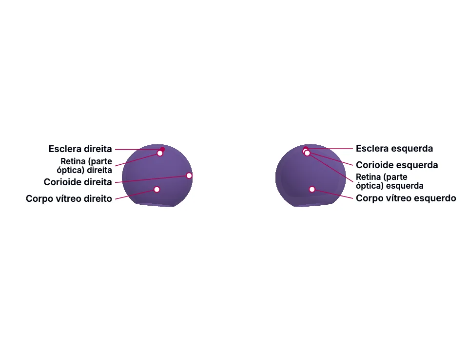
A parede do globo ocular tem três camadas concêntricas: 'the wall of the eyeball is composed of three layers: the fibrous tunic, the vascular tunic, and the retina.' A túnica fibrosa é a mais externa - 'the fibrous tunic is the outermost layer and consists of the anterior cornea and the posterior sclera' -, e sua porção posterior, a esclera, 'is the white of the eye and covers the entire eyeball except for the cornea. It protects its inner parts, gives shape to the eyeball, and makes it more rigid.' A túnica vascular, ou úvea, é a camada média: 'the vascular tunic, also known as the uvea, is the middle layer of the eyeball.' Sua porção posterior é a corioide, que reveste a face interna da esclera: 'the choroid is the posterior portion of the vascular tunic, and it lines the internal surface of the sclera. Its numerous blood vessels provide nutrients to the retina.' A retina é a terceira e mais interna: 'the third and innermost layer of the eyeball is the retina.'
É exatamente por estar encaixada entre a corioide (por fora) e o vítreo (por dentro) que a retina depende da corioide para se manter viva: a corioide 'forms the posterior portion of the uveal tract, and nourishes the outer retinal layers', e os nutrientes que atravessam o choriocapilar, logo atrás do epitélio pigmentar, sustentam a camada de fotorreceptores - 'nutrients from the choriocapillaris behind the RPE support the photoreceptor layer. The photoreceptors and most of the OPL rely indirectly on the choriocapillaris.' Essa dependência é ainda mais absoluta num único ponto: a fóvea não tem vaso sanguíneo retiniano próprio, e depende inteiramente da corioide para seu oxigênio - 'there are no retinal blood vessels within the fovea, and retinal tissue within this area depends entirely on the choroid for its oxygen requirements.'
A cavidade vítrea fica entre o cristalino e a retina, e é preenchida pelo corpo vítreo - uma substância gelatinosa transparente que mantém a retina fisicamente encostada contra a corioide: 'the larger cavity of the eyeball is the vitreous cavity, located between the lens and the retina. Within the vitreous chamber is the vitreous body, a transparent jellylike substance that holds the retina against the choroid.' Com a idade, essa relação muda: o descolamento posterior do vítreo (DPV) é a separação entre o córtex vítreo posterior e a membrana limitante interna da retina - 'posterior vitreous detachment (PVD) is the separation of the posterior vitreous cortex and the internal limiting membrane of the retina and is the most common cause of floaters.'
Na maioria das vezes essa separação é benigna (a causa mais comum de moscas volantes), mas quando é ANÔMALA ela produz tração na própria interface que antes segurava a retina no lugar: 'anomalous PVD can lead to various deleterious effects on the retina as well as vitreous as a result of abnormal traction at the vitreoretinal interface.' É exatamente essa tração, atuando sobre um ponto fraco da retina periférica, que a literatura de descolamento de retina liga ao início do quadro mais agressivo: 'a superior horseshoe tear can cause rapid progression of superior RRD that involves the fovea early in an older patient with the posterior vitreous detachment (PVD) and liquefied vitreous.'
A retina tem uma camada mais externa, o epitélio pigmentar (EPR), encostada na corioide, e uma mais interna, a membrana limitante interna (MLI), voltada para a cavidade vítrea: 'the outermost layer called the retinal pigment epithelium (RPE), abuts the choroid, and the innermost layer, or the internal limiting membrane (ILM), faces the vitreous cavity.' O descolamento de retina é, por definição, a separação entre a retina neurossensorial e esse EPR subjacente: 'retinal detachment (RD) occurs when the neurosensory retina separates from the underlying RPE.'
No tipo regmatogênico - o mais comum -, essa separação acontece porque líquido passa da cavidade vítrea, através de uma ruptura ou quebra retiniana, para o espaço potencial entre a retina sensorial e o EPR: 'rhegmatogenous RDs are the most common, caused by fluid passing from the vitreous cavity via a retinal tear or a break into the potential space between the sensory retina and the RPE (subretinal space).' Isso é inteiramente diferente do tipo tracional, que NÃO exige ruptura nenhuma: ali, membranas proliferativas se contraem e elevam a retina mecanicamente - 'tractional detachments occur when proliferative membranes contract and elevate the retina.'
In the clinic
Essa dependência estrutural é o que torna o descolamento de retina uma emergência: uma vez que a retina neurossensorial se separa da corioide, 'it loses its oxygen and nutrient supply, leading to the death of the tissue.' E porque a fóvea não tem rede vascular própria, é a estrutura mais vulnerável a esse desfecho - 'a detachment of the clinical macula (or anatomical fovea) can lead to permanent damage to the photoreceptors in this location': é por isso que o status da mácula (descolada ou poupada) é o fator que mais pesa no prognóstico visual de um descolamento de retina.
Uma vez que a tração do DPV abre uma ruptura de espessura total na retina, o próprio vítreo liquefeito - que antes protegia a retina, mantendo-a encostada - passa a ser o que a separa: 'this process is facilitated by traction over the retinal break that keeps the gap open, ensuring that the entry of liquefied vitreous or fluid to the subretinal space supersedes the RPE pump, which helps absorb the subretinal fluid.' Por isso um descolamento de retina regmatogênico não nasce de uma causa só: precisa de uma ruptura de espessura total, de vítreo liquefeito, com ou sem tração adicional sobre a ruptura - 'for an RRD to occur, the following are needed: A full-thickness retinal break Liquified vitreous, with or without Traction over the break'
Qualquer que seja o mecanismo, o resultado anatômico é o mesmo: a retina perde o suprimento que só a corioide, através do EPR, dava. E o achado mais decisivo para o prognóstico visual é justamente o status da mácula: 'one study's findings showed that 83% of patients who still had the macula attached had a best-corrected visual acuity of 20/40 or better', enquanto 'if the macula is detached, however, the visual prognosis is relatively poor' - a mesma fóvea sem vaso próprio, e por isso mais dependente da corioide, que o primeiro bloco já havia identificado.
Open the deeper layer
Going deeper
O mesênquima extracelular que envolve o olho em desenvolvimento - formado por células da crista neural que MIGRAM até ali, mais mesoderma: 'the eye is derived from the neuroepithelium, the surface ectoderm, and the extracellular mesenchyme which consists of both the neural crest and mesoderm.'
Esclera - origina-se do mesênquima extracelular: 'the extracellular mesenchyme forms the sclera, the corneal endothelium, stroma, blood vessels, muscles, and vitreous.'; Corpo vítreo - mesma origem mesenquimal da esclera, citada na MESMA frase da fonte acima.; Melanócitos da corioide - não vêm do mesênquima extracelular em bloco, mas especificamente de células da crista neural que migram até o olho em formação: 'neural crest cells migrate toward the developing eye and differentiate into choroidal melanocytes, which provide pigment that reduces light scatter in the mature retina.' (fonte independente, Jozsa & Hall.); Retina (camada pigmentada) - a EXCEÇÃO: ao contrário da esclera, do vítreo e dos melanócitos da corioide, que chegam ao olho por migração (crista neural/mesênquima), a camada pigmentada da retina se forma NO LOCAL, direto do neuroepitélio óptico: 'the outer pigmented layer arises directly from the optic neuroepithelium.' A retina inteira, aliás, tem dupla origem germinativa - ectodérmica e de crista neural: 'the retina is derived from ectodermal and neural crest embryonic cells' -, e é a camada neuroblástica interna, não a pigmentada, que dá origem aos fotorreceptores: 'the internal neuroblastic layer gives rise to light-sensing rods and cones.'
Test yourself before moving on
Quais são as três túnicas da parede do globo ocular, e qual delas é a mais interna?
The answer stays in Assessment, with no spoiler here.
Answer in AssessmentSources (4)
- Rehman I, Hazhirkarzar B, Patel BC. Anatomy, Head and Neck, Eye. [Updated 2023 Jul 24]. In: StatPearls [Internet]. Treasure Island (FL): StatPearls Publishing; 2026 Jan-.
Anatomically, the wall of the eyeball is composed of three layers: the fibrous tunic, the vascular tunic, and the retina.
- Jozsa F, Hall WA. Neuroanatomy, Retina. [Updated 2026 Feb 22]. In: StatPearls [Internet]. Treasure Island (FL): StatPearls Publishing; 2026 Jan-.
The choroid, forming the posterior portion of the uveal tract, nourishes the outer retinal layers.
- Blair K, Czyz CN. Retinal Detachment. [Updated 2024 Feb 12]. In: StatPearls [Internet]. Treasure Island (FL): StatPearls Publishing; 2026 Jan-.
There are no retinal blood vessels within the fovea, and retinal tissue within this area depends entirely on the choroid for its oxygen requirements.
- Ahmed F, Tripathy K. Posterior Vitreous Detachment. [Updated 2023 Aug 25]. In: StatPearls [Internet]. Treasure Island (FL): StatPearls Publishing; 2026 Jan-.
Posterior vitreous detachment (PVD) is the separation of the posterior vitreous cortex and the internal limiting membrane of the retina and is the most common cause of floaters.
Where this shows up in practice
1 clinical case in the library requires this topic. See where it decides the management.
Glaucoma agudo de ângulo fechado: a dor de cabeça que não é enxaqueca
Medicina de emergência
Open the case for free
More Anatomia summaries
After reading, use what you learned
Atlas
See the structures of the topic in 3D and move through the layers.
Clinical case
Apply the content to an on-call decision.
Exam
Read the result that closes the reasoning.
Assessment
Answer the block questions and see where you stand.
AI tutor
Ask about what the text did not settle.
Start free: create an account and try a sample of the 3D Atlas, the clinical case, the exam, the assessment and the AI tutor. No credit card.
Start free