Quadríceps e isquiotibiais: reflexo patelar, ruptura do tendão do quadríceps e lesão muscular no esporte
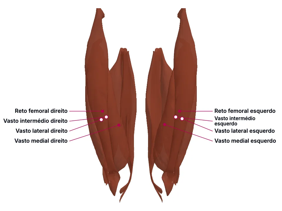
Os quatro componentes do quadríceps convergem num tendão único que continua como ligamento patelar até a tuberosidade da tíbia, e é esse arco que o reflexo patelar testa - raiz L2-L4, nervo femoral, logo abaixo da patela -, com o mesmo estiramento do fuso muscular que contrai o quadríceps inibindo reciprocamente o isquiotibial, de modo que hiporreflexia sugere lesão de neurônio motor inferior nesse arco e hiperreflexia sugere lesão de neurônio motor superior. O reto femoral se origina na espinha ilíaca anterior inferior - diferente dos três vastos, originados no próprio fêmur -, o que lhe dá uma ação extra de flexão do quadril e explica por que é a porção do quadríceps mais envolvida em lesão muscular indireta, 60% dos casos na área proximal; já no trauma agudo do mecanismo extensor do joelho, um defeito palpável suprapatelar indica ruptura do tendão do quadríceps, enquanto um defeito palpável na própria patela indica fratura de patela. Os isquiotibiais compartilham origem no túber isquiático - semitendíneo e a cabeça longa do bíceps femoral no mesmo tendão conjunto, semimembranáceo em origem SEPARADA, mais lateral -, exceto a cabeça curta do bíceps femoral, que se origina no próprio fêmur e por isso só cruza o joelho, não o quadril; essa mesma cabeça recebe inervação do nervo fibular comum na bifurcação do nervo isquiático, diferente das outras três (nervo tibial), o que explica a estimulação assíncrona por trás de o bíceps femoral ser o isquiotibial mais frequentemente lesionado num grupo muscular cuja lesão já está entre as mais comuns no esporte.
Educational material written with AI support and checked by automated review, without a named medical reviewer. It does not replace the textbook or clinical guidance.
Essential
Os quatro componentes do quadríceps convergem num tendão único: 'The four heads are directed downward to form a single tendon, the quadriceps muscle tendon, which fits on the patella.' - confirmado por uma segunda fonte independente: 'The quadriceps femoris connects to the patella via the quadriceps tendon, which then extends to insert at the tuberosity of the tibia.' As fibras mais superficiais desse tendão continuam sobre a patela e se inserem na tuberosidade da tíbia como ligamento patelar: 'The most superficial fibers of the tendon continue and cover the patella, inserting on the tibial tuberosity called the patellar ligament.'
É esse arco inteiro que o reflexo patelar testa: 'Knee (quadriceps, patellar): L2-L4 (predominately L4), femoral, just inferior to the patella' O mecanismo é o estiramento do fuso muscular, cujo impulso aferente sinapsa direto num neurônio motor: 'This activates stretch receptors in the muscle spindle, of which the cell bodies are in the dorsal root ganglia, and an afferent impulse is conducted via these Ia afferent neurons to the spinal cord where they directly synapse on an alpha motor neuron.' Um ramo do MESMO sinal ativa um interneurônio inibitório que impede a contração do grupo antagonista: 'When an MSR is initiated there is also a signal that branches off from the Ia afferent stretch neuron and activates an inhibitory interneuron that will prevent the opposing (flexor or extensor) muscle group from activating during the reflex.' - no joelho, isso significa inibir o isquiotibial enquanto o quadríceps contrai: 'at the knee, the quadriceps opposes the hamstring, so when the knee jerk reflex is elicited it inhibits the hamstring motor neurons while the quadriceps are stimulated to contract.'
O reto femoral se origina na espinha ilíaca anterior inferior: 'the rectus femoris (RS) that originates from the anterior inferior iliac spine with the direct tendon and the upper rim of the acetabulum with the indirect tendon.' Os três vastos, ao contrário, se originam no próprio corpo do fêmur - o vasto lateral na face lateral do grande trocânter e na linha áspera: 'The vastus lateralis (VL) originates from the lateral face of the great trochanter, the gluteal tuberosity, and the lateral lip of the linea aspera.' o vasto medial no colo anatômico do fêmur e na linha áspera: 'The vastus medialis (VM) runs from the anatomical neck of the femur and the medial lip of the linea aspera.' e o vasto intermédio no próprio corpo do fêmur: 'The vastus intermedius (VI) originates from the proximal three-fourths of the anterior and lateral faces of the femoral body and the lateral lip of the linea aspera.'
Essa origem diferente dá ao reto femoral uma ação extra: 'All 4 function to extend the knee, but the rectus femoris is also an accessory flexor of the hip.' - confirmado por uma segunda fonte independente: 'The rectus femoris is responsible for thigh flexion at the hip and knee extension.' enquanto os vastos ficam limitados à extensão do joelho: 'The vastus muscles are responsible for knee extension and stabilization of the patella.'
O tendão do quadríceps, a patela e o ligamento patelar formam, juntos, o mecanismo extensor do joelho: 'The quadriceps tendon, in combination with the patellar tendon and the patella bone, makes up the extensor mechanism of the lower leg.' A patela, especificamente, é uma peça desse mesmo mecanismo: 'Patellar fractures are clinically significant injuries that may compromise the extensor mechanism, disrupt articular congruity, and impair long-term knee function.'
Quando o tendão do quadríceps se rompe, o mecanismo extensor fica comprometido de um jeito característico: 'The clinical diagnostic triad includes acute pain, loss of active straight leg raising, and a palpable suprapatellar defect.'
O semitendíneo e a cabeça longa do bíceps femoral compartilham um tendão conjunto na face súpero-medial do túber isquiático: 'The common (conjoint) tendon from the superior medial quadrant of the posterior ischial tuberosity (with biceps femoris long head)' O semimembranáceo tem origem SEPARADA desse tendão conjunto, na face súpero-lateral do túber isquiático: 'Superior lateral aspect of the ischial tuberosity' - 'One variation noted in current research is the distinct origin of the biceps femoris long head and semitendinosus from separate tendons attached to the ischial tuberosity, as opposed to their more common shared origin from the common (conjoint) tendon of the ischial tuberosity.' E a cabeça CURTA do bíceps femoral é a exceção maior entre todos os isquiotibiais - origina-se no próprio fêmur, não no túber isquiático: 'Originating at the pelvis and running posteriorly along the femur, most muscles of the hamstring complex cross both the femoroacetabular and tibiofemoral joints. The short head of the biceps femoris is an exception, arising from the lateral lip of the femoral linea aspera, distal to the femoroacetabular joint.'
Consequência dessa exceção de origem: 'All the hamstring muscles traverse two joints (hip and knee) from origin to insertion except the short head of biceps femoris, which only traverses knee joint from its origin to insertion.'
In the clinic
Como o arco tem componentes de neurônio motor inferior (nervo periférico, segmento medular) e superior (trato corticoespinal descendente), a alteração do reflexo aponta o nível da lesão: 'hyperreflexia can indicate an upper motor neuron lesion, whereas hyporeflexia can indicate a lower motor neuron lesion.'
Consequência clínica, da MESMA fonte da anatomia do reto femoral: 'The rectus femoris is the muscle portion most involved in indirect lesions, and in 60% of patients, the trauma involves the most proximal area'
Quando é a patela que fratura, o defeito palpável aparece NA PRÓPRIA patela, não acima dela: 'Palpation of closed injuries may reveal crepitus, a palpable fracture gap, and swelling indicative of hemarthrosis.' e o exame usa o mesmo teste de elevação da perna esticada para avaliar o mecanismo extensor: 'A straight leg raise test must be performed to assess the extensor mechanism, although pain may limit the examination.' - mesmo mecanismo comprometido nos dois quadros, defeito palpável em lugar diferente.
O bíceps femoral é o isquiotibial mais frequentemente lesionado: 'The biceps femoris is the most frequently injured hamstring muscle, followed by the semimembranosus and semitendinosus.' e a PRÓPRIA fonte da lesão atribui isso à dupla inervação de suas duas cabeças: 'dual nerve supply of two heads of biceps femoris leading to asynchronous stimulation as well as an anatomical variance of attachment of its two heads makes it more commonly injured hamstring.' A lesão de isquiotibial, de modo geral, está entre as mais comuns no esporte: 'Hamstring strains are among the most common injuries in both elite and recreational athletes, with a high recurrence rate.'
Open the deeper layer
Going deeper
O nervo isquiático bifurca, ao nível da articulação tibiofemoral (perto do joelho), em dois ramos: 'At the level of the tibiofemoral joint, the sciatic nerve bifurcates into the tibial and common fibular (peroneal) nerves.'
Semimembranáceo - recebe o ramo TIBIAL: 'The tibial nerve supplies the semimembranosus, semitendinosus, and long head of the biceps femoris, while the common fibular nerve innervates the short head of the biceps femoris' Mesmo músculo cuja origem é SEPARADA do tendão conjunto que a cabeça longa do bíceps femoral compartilha com o semitendíneo: 'One variation noted in current research is the distinct origin of the biceps femoris long head and semitendinosus from separate tendons attached to the ischial tuberosity, as opposed to their more common shared origin from the common (conjoint) tendon of the ischial tuberosity.' - originando-se, em vez disso, na face súpero-lateral do túber isquiático: 'Superior lateral aspect of the ischial tuberosity'; Semitendíneo - também recebe o ramo TIBIAL: 'The tibial nerve supplies the semimembranosus, semitendinosus, and long head of the biceps femoris, while the common fibular nerve innervates the short head of the biceps femoris' Origina-se no mesmo tendão conjunto da cabeça longa do bíceps femoral, na face súpero-medial do túber isquiático: 'The common (conjoint) tendon from the superior medial quadrant of the posterior ischial tuberosity (with biceps femoris long head)' Cruza tanto o quadril quanto o joelho, igual à maioria dos isquiotibiais: 'Originating at the pelvis and running posteriorly along the femur, most muscles of the hamstring complex cross both the femoroacetabular and tibiofemoral joints. The short head of the biceps femoris is an exception, arising from the lateral lip of the femoral linea aspera, distal to the femoroacetabular joint.'; Cabeça LONGA do bíceps femoral - também recebe o ramo TIBIAL: 'The tibial nerve supplies the semimembranosus, semitendinosus, and long head of the biceps femoris, while the common fibular nerve innervates the short head of the biceps femoris'; Cabeça CURTA do bíceps femoral - recebe o ramo FIBULAR COMUM, diferente das outras três: 'The tibial nerve supplies the semimembranosus, semitendinosus, and long head of the biceps femoris, while the common fibular nerve innervates the short head of the biceps femoris' A MESMA cabeça que tem origem diferente (fêmur, não túber isquiático, distal à articulação do quadril: 'Originating at the pelvis and running posteriorly along the femur, most muscles of the hamstring complex cross both the femoroacetabular and tibiofemoral joints. The short head of the biceps femoris is an exception, arising from the lateral lip of the femoral linea aspera, distal to the femoroacetabular joint.') também tem inervação diferente - a dupla divergência que a própria fonte da lesão liga à estimulação assíncrona entre as duas cabeças do bíceps femoral: 'dual nerve supply of two heads of biceps femoris leading to asynchronous stimulation as well as an anatomical variance of attachment of its two heads makes it more commonly injured hamstring.'
Test yourself before moving on
Ao testar o reflexo patelar de um paciente, o examinador nota resposta ausente (arreflexia). Que raiz nervosa e que nervo periférico compõem esse arco reflexo, e por que é o quadríceps - não o isquiotibial - que contrai quando o martelo bate no tendão patelar?
The answer stays in Assessment, with no spoiler here.
Answer in AssessmentSources (9)
- Bordoni B, Varacallo MA. Anatomy, Bony Pelvis and Lower Limb: Thigh Quadriceps Muscle. [Updated 2023 May 8]. In: StatPearls [Internet]. Treasure Island (FL): StatPearls Publishing; 2026 Jan-.
The four heads are directed downward to form a single tendon, the quadriceps muscle tendon, which fits on the patella.
- Khan A, Arain A. Anatomy, Bony Pelvis and Lower Limb: Anterior Thigh Muscles. [Updated 2023 Mar 17]. In: StatPearls [Internet]. Treasure Island (FL): StatPearls Publishing; 2026 Jan-.
The quadriceps femoris connects to the patella via the quadriceps tendon, which then extends to insert at the tuberosity of the tibia.
- Zimmerman B, Hubbard JB. Deep Tendon Reflexes. [Updated 2023 Jul 24]. In: StatPearls [Internet]. Treasure Island (FL): StatPearls Publishing; 2026 Jan-.
Knee (quadriceps, patellar): L2-L4 (predominately L4), femoral, just inferior to the patella
- Attum B, Varacallo MA. Anatomy, Bony Pelvis and Lower Limb: Thigh Muscles. [Updated 2022 Dec 17]. In: StatPearls [Internet]. Treasure Island (FL): StatPearls Publishing; 2026 Jan-.
All 4 function to extend the knee, but the rectus femoris is also an accessory flexor of the hip.
- Pope JD, El Bitar Y, Mabrouk A, et al. Quadriceps Tendon Rupture. [Updated 2023 Apr 22]. In: StatPearls [Internet]. Treasure Island (FL): StatPearls Publishing; 2026 Jan-.
The quadriceps tendon, in combination with the patellar tendon and the patella bone, makes up the extensor mechanism of the lower leg.
- Mabrouk A, Pilson H. Patella Fractures. [Updated 2026 Aug 15]. In: StatPearls [Internet]. Treasure Island (FL): StatPearls Publishing; 2026 Jan-.
Patellar fractures are clinically significant injuries that may compromise the extensor mechanism, disrupt articular congruity, and impair long-term knee function.
- Vaughn JE, Cohen-Levy WB. Anatomy, Bony Pelvis and Lower Limb: Posterior Thigh Muscles. [Updated 2023 Aug 21]. In: StatPearls [Internet]. Treasure Island (FL): StatPearls Publishing; 2026 Jan-.
The common (conjoint) tendon from the superior medial quadrant of the posterior ischial tuberosity (with biceps femoris long head)
- Hu Y, Raja A. Anatomy, Bony Pelvis and Lower Limb, Hamstring Muscle. [Updated 2026 Jan 31]. In: StatPearls [Internet]. Treasure Island (FL): StatPearls Publishing; 2026 Jan-.
Originating at the pelvis and running posteriorly along the femur, most muscles of the hamstring complex cross both the femoroacetabular and tibiofemoral joints. The short head of the biceps femoris is an exception, arising from the lateral lip of the femoral linea aspera, distal to the femoroacetab…
- Poudel B, Pandey S. Hamstring Injury. [Updated 2023 Aug 8]. In: StatPearls [Internet]. Treasure Island (FL): StatPearls Publishing; 2026 Jan-.
All the hamstring muscles traverse two joints (hip and knee) from origin to insertion except the short head of biceps femoris, which only traverses knee joint from its origin to insertion.
More Anatomia summaries
After reading, use what you learned
Atlas
See the structures of the topic in 3D and move through the layers.
Clinical case
Apply the content to an on-call decision.
Exam
Read the result that closes the reasoning.
Assessment
Answer the block questions and see where you stand.
AI tutor
Ask about what the text did not settle.
Start free: create an account and try a sample of the 3D Atlas, the clinical case, the exam, the assessment and the AI tutor. No credit card.
Start free