Pulmões e vias aéreas: da traqueia ao segmento broncopulmonar
In 30 seconds
A traqueia se bifurca na carina em dois brônquios principais assimétricos — o direito mais largo, curto e vertical; o esquerdo mais estreito, longo e horizontal — e essa assimetria decide para onde vai um corpo estranho aspirado.
Scope and method of this summary
A traqueia se bifurca na carina em dois brônquios principais assimétricos — o direito mais largo, curto e vertical; o esquerdo mais estreito, longo e horizontal — e essa assimetria decide para onde vai um corpo estranho aspirado. Cada brônquio se ramifica em lobares e depois em segmentares, e cada segmento broncopulmonar, por ter brônquio próprio, é uma unidade que a cirurgia pode isolar sem tocar no resto do lobo. Por fora de tudo isso, a pleura parietal (que dói) e a visceral (que não dói) explicam por que só certas doenças pleurais doem, e por que o ar ou o líquido no espaço entre elas muda o exame físico do jeito que muda.
Educational material written with AI support and checked by automated review, without a named medical reviewer. It does not replace the textbook or clinical guidance.
Essential
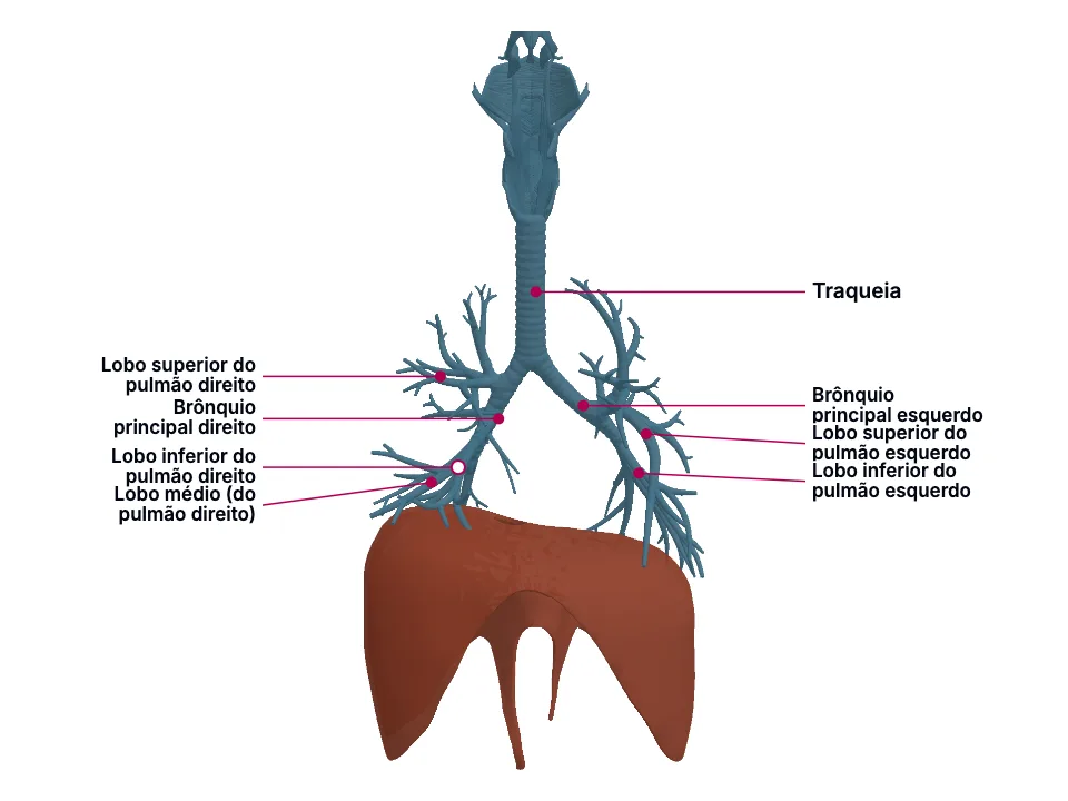
A traqueia termina na carina, o ponto de bifurcação nos dois brônquios principais ('terminates at the bifurcation into the primary bronchi at the carina'). Ali, os dois brônquios não são espelhados: o direito é mais largo, mais curto e mais vertical, enquanto o esquerdo é mais estreito, mais longo e mais horizontal, porque precisa contornar o arco da aorta — 'the right main bronchus has a wider diameter, is shorter, and lies more vertically relative to the hilum' contra 'the left main bronchus has a smaller diameter and is more horizontal; it must pass inferior to the arch of the aorta and anterior to the esophagus and thoracic aorta to reach the left lung's hilum'.
Um objeto aspirado desce pela via de MENOR resistência ao seu próprio peso e trajetória: o brônquio mais largo, mais curto e mais alinhado ao eixo da traqueia (o direito) recebe o fluxo de preferência sobre o mais estreito e mais desviado (o esquerdo, que precisa curvar sob a aorta). Não é uma diferença de calibre isolada — é largura, comprimento E ângulo, os três a favor do lado direito ao mesmo tempo.
Duas fissuras dividem o pulmão direito em três lobos — 'the right lung is comprised of the right upper (RUL), middle (RML), and lower (RLL) lobes' e 'oblique and horizontal fissures separate the right lung's 3 lobes' —, enquanto o esquerdo tem só uma fissura e dois lobos — 'the left lung consists of the left upper (LUL) and lower (LLL) lobes' —, porque cede espaço a 'a cardiac notch—a concavity accommodating the left cardiac ventricle—in the left lung'. Dentro de cada lobo, brônquios segmentares de 3ª ordem — 'segmental bronchi are the 3rd-order branches off the 2nd-order branches (lobar bronchi) that arise from the main bronchus' — ventilam cada um o seu próprio segmento broncopulmonar: 'each segmental bronchi supply a bronchopulmonary segment, which are the largest subdivisions of the lobe'.
Como cada segmento broncopulmonar tem brônquio segmentar PRÓPRIO — não compartilhado com o segmento vizinho —, ele é uma unidade estruturalmente separável dentro do lobo: dá para interromper o suprimento de ar de UM segmento sem tocar nos outros. É essa independência de via aérea, segmento a segmento, que torna o segmento (e não só o lobo) uma unidade que a cirurgia pode isolar.
A pleura tem duas camadas: 'the outer layer is called the parietal pleura and attaches to the chest wall. The inner layer is called the visceral pleura and covers the lungs, blood vessels, nerves, and bronchi.' Só a parietal recebe nervo somático: 'the costal and cervical portions of the parietal pleura are innervated by the intercostal nerve, and the diaphragmatic portion is supplied by the phrenic nerve.'
'The parietal pleura is the only portion of the pleura that can sense pain. The visceral pleura receives its nerve supply via the autonomic nervous system (ANS) and lacks sensory innervation.' Ou seja: um processo que fica restrito ao pulmão e à pleura visceral (pneumonia, por exemplo) não tem, por si só, a inervação somática que produz dor localizada — a dor só aparece quando o processo alcança ou irrita a pleura PARIETAL, que é a camada com nervo intercostal (parede costal) ou frênico (face diafragmática).
In the clinic
O achado é direto na fonte: 'the right lung is more commonly affected by inhaled foreign bodies', porque 'aspirated foreign objects are more likely to settle in the right bronchus'. A posição do paciente no momento da aspiração decide até o lobo exato: 'while lying supine, the foreign body usually enters the right lower lobe (superior portion). While lying on the right side, the foreign body usually enters the right upper lobe. While upright, the foreign object usually enters the right lower lobe (lower portion)'.
A fonte nomeia essa distinção operacional direto: 'a lobectomy is the removal of an entire lung lobe, while a segmentectomy is the excision of a lung segment' — e 'a segmentectomy may be performed on benign segment masses to preserve the lung', preservando os demais segmentos daquele mesmo lobo justamente porque cada um tinha brônquio próprio para começar.
O pneumotórax mostra a mesma lógica pelo lado mecânico: o ar que entra no espaço pleural dissolve a tensão superficial que colava as duas camadas — 'when air enters the pleural space, the surface tension will disappear' —, o pulmão se afasta da parede torácica e o exame muda por inteiro: 'percussion and auscultation will reveal a hyper-resonant chest with no breath sounds'. E quando o que se acumula no espaço pleural é líquido, não ar, ele desce com a gravidade até o ponto mais baixo: 'pleural effusions usually collect in the costodiaphragmatic recess' — o mesmo recesso cuja porção diafragmática é inervada pelo frênico, citado acima.
Open the deeper layer
Going deeper
O divertículo respiratório (broto pulmonar), um crescimento ventral do endoderma do intestino anterior (foregut): 'the first traces of the bronchial tree appear as the respiratory diverticulum (lung bud), which is located ventrally on the foregut as an outgrowth.'
Septo traqueoesofágico — duas cristas longitudinais que crescem dos dois lados do broto e se fundem, separando o esôfago da traqueia primitiva em desenvolvimento: 'these are two longitudinal ridges on either side of the bud that fuse to form the tracheoesophageal septum, thus dividing the esophagus from the developing primitive trachea (lung bud).'; Traqueia — a porção proximal do próprio divertículo, que permanece como conduto único depois da separação do esôfago.; Brônquios principais direito e esquerdo — os brotos brônquicos bilaterais do divertículo, que aumentam de tamanho no início da 5ª semana: 'the left and right main bronchi (from the bronchial buds) enlarge at the beginning of the fifth week.'; Brônquios lobares e segmentares — gerações sucessivas de ramificação dicotômica dos brotos brônquicos, que já na vida fetal produzem o número de brônquios terciários que a anatomia madura preserva por lado: 'these secondary bronchi subdivide in a dichotomous fashion, thus forming eight tertiary bronchi in the left lung and ten tertiary bronchi in the right' — o mesmo padrão assimétrico (mais à direita que à esquerda) que aparece nos segmentos broncopulmonares do pulmão adulto.
Test yourself before moving on
Uma criança aspira um pedaço de amendoim enquanto está em pé. Pelo que as fontes desta unidade descrevem sobre a anatomia dos brônquios principais, em qual pulmão e em qual região dele o amendoim tem mais chance de parar?
The answer stays in Assessment, with no spoiler here.
Answer in AssessmentSources (4)
- Derricott H, Jimenez J, La Rose J. Anatomy, Head and Neck, Trachea. [Updated 2026 Jun 8]. In: StatPearls [Internet]. Treasure Island (FL): StatPearls Publishing; 2026 Jan-.
terminates at the bifurcation into the primary bronchi at the carina at the level of the 4th to 5th thoracic vertebrae
- Amador C, Weber C, Varacallo MA. Anatomy, Thorax, Bronchial. [Updated 2023 Aug 8]. In: StatPearls [Internet]. Treasure Island (FL): StatPearls Publishing; 2026 Jan-.
The right main bronchus has a wider diameter, is shorter, and lies more vertically relative to the hilum.
- Chaudhry R, Omole AE, Bordoni B. Anatomy, Thorax, Lungs. [Updated 2024 Apr 20]. In: StatPearls [Internet]. Treasure Island (FL): StatPearls Publishing; 2026 Jan-.
The right lung is comprised of the right upper (RUL), middle (RML), and lower (RLL) lobes.
- Mahabadi N, Goizueta AA, Bordoni B. Anatomy, Thorax, Lung Pleura And Mediastinum. [Updated 2024 Mar 24]. In: StatPearls [Internet]. Treasure Island (FL): StatPearls Publishing; 2026 Jan-.
The outer layer is called the parietal pleura and attaches to the chest wall. The inner layer is called the visceral pleura and covers the lungs, blood vessels, nerves, and bronchi.
Where this shows up in practice
1 clinical case in the library requires this topic. See where it decides the management.
Febre, tosse e falta de ar com opacidades na tomografia de tórax
Radiologia e diagnóstico por imagem
Open the case for free
More Anatomia summaries
After reading, use what you learned
Atlas
See the structures of the topic in 3D and move through the layers.
Clinical case
Apply the content to an on-call decision.
Exam
Read the result that closes the reasoning.
Assessment
Answer the block questions and see where you stand.
AI tutor
Ask about what the text did not settle.
Start free: create an account and try a sample of the 3D Atlas, the clinical case, the exam, the assessment and the AI tutor. No credit card.
Start free