Nervos oculares: a via pupilar e os nervos motores do olho
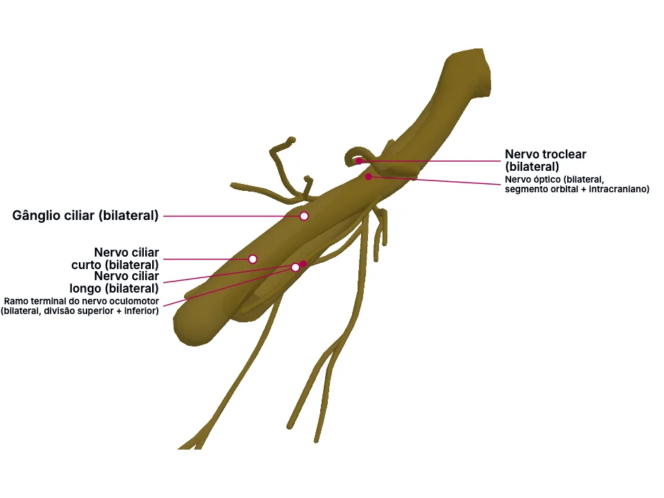
O reflexo fotomotor fecha um arco entre o nervo óptico (aferência) e o nervo oculomotor, que sinapsa no gânglio ciliar antes de seguir pelo nervo ciliar curto até o esfíncter da pupila (eferência) - o arco testado pelo defeito pupilar aferente relativo -, e a estratificação das fibras dentro do próprio nervo oculomotor explica por que uma compressão externa (aneurisma de comunicante posterior) dilata a pupila enquanto uma microangiopatia (diabetes) a poupa. A via simpática ocular percorre o caminho oposto - três neurônios até o nervo ciliar LONGO -, cuja interrupção produz a tríade da síndrome de Horner (ptose parcial, miose, anidrose facial), e o nervo troclear - único nervo craniano motor dorsal e decussante - inerva sozinho o oblíquo superior, cuja paralisia produz diplopia vertical pior no olhar para baixo. A pupila de Argyll-Robertson, dissociação luz-perto da neurossífilis, ilustra como uma lesão mesencefálica dorsal bem localizada pode poupar a via, ligeiramente mais ventral, da acomodação.
Educational material written with AI support and checked by automated review, without a named medical reviewer. It does not replace the textbook or clinical guidance.
Essential
A perna AFERENTE do reflexo fotomotor começa na retina: 'Photoreceptor cells in the outer layers of the retina, which are called rods and cones, convert light stimuli into neuronal impulses. These signals are then relayed to the bipolar cells, which interact with ganglion cells, which in turn coalesce to form the optic disc and optic nerve (CN II).'
A perna EFERENTE segue pelo nervo oculomotor (III), faz sinapse no gânglio ciliar, e termina pelo nervo ciliar CURTO no esfíncter da pupila: 'Efferent parasympathetic preganglionic fibers travel on the oculomotor nerve and synapse with the ciliary ganglion, which sends postganglionic axons to directly innervate the iris sphincter muscles. The contraction of the iris sphincter muscles leads to pupillary constriction (miosis).' - fechando o arco que contrai a pupila em resposta à luz.
O nervo oculomotor (III par) tem dois componentes funcionais em camadas diferentes do mesmo tronco: 'The third cranial nerve, also known as the oculomotor nerve, contains 2 major functional components. Parasympathetic fibers, located superficially, innervate the ciliary muscle and sphincter pupillae, mediating lens accommodation and pupillary constriction. Somatic motor fibers, located deeper, innervate the levator palpebrae superioris, which elevates the upper eyelid, and 4 extraocular muscles—the superior, medial, and inferior recti, as well as the inferior oblique.' As fibras pupilares recebem irrigação diferente das fibras motoras somáticas mais profundas: 'Before orbital entry, fibers responsible for pupillary constriction are located superficially within the nerve trunk and receive vascular supply from pial blood vessels. In contrast, deeper somatic motor fibers are supplied by the vasa vasorum.'
Uma massa que comprime o nervo DE FORA para dentro - o aneurisma de comunicante posterior é o exemplo clássico - atinge primeiro a camada superficial: 'Intracranial aneurysm is the most concerning cause of acute cranial nerve III palsy and typically results from external compression by an enlarging aneurysm. The posterior communicating artery constitutes the classic site, although aneurysms of the internal carotid and basilar arteries may also produce cranial nerve III palsy.' Já uma microangiopatia - o diabetes é o exemplo clássico - afeta o interior do tronco nervoso, poupando a camada pupilar: 'Microvascular ischemia, often termed microvascular palsy or diabetic cranial nerve III palsy , is the most common etiology in adults. Small-vessel disease underlies most cases, with hypertension, advancing age, and other vascular risk factors increasing susceptibility.' O mecanismo que separa as duas apresentações: 'Pathologies such as aneurysms, uncal herniation, or tumors compress the nerve externally and preferentially involve the superficial pupillomotor fibers and associated blood supply. In contrast, medical causes such as diabetes mellitus or hypertension-related microangiopathy affect the vasa vasorum and typically spare the pupillary fibers, resulting in pupil-sparing cranial nerve III palsy.'
A via simpática ocular é um trajeto de três neurônios, do hipotálamo até os nervos ciliares LONGOS: 'The nerve supply consists of a 3-neuron pathway extending from the posterolateral hypothalamus to the long ciliary nerves, which innervate the iris dilator and superior tarsal muscle, also known as the Müller muscle.'
Depois de fazer sinapse no gânglio cervical superior, as fibras pós-ganglionares entram na órbita pela fissura orbital superior, acompanhando a divisão oftálmica do trigêmeo, e seguem como nervo ciliar longo: 'the fibers enter the orbit through the superior orbital fissure and travel with the ophthalmic division of the trigeminal nerve (cranial nerve V) before continuing as the long ciliary nerves.' - o mesmo par de nervos ciliares da via pupilar parassimpática, mas do lado simpático, terminando no dilatador da pupila, não no esfíncter.
O nervo troclear (IV par) é o único nervo craniano motor que emerge do dorso do tronco encefálico e decussa antes de chegar ao músculo que inerva: 'arises from the midbrain at the level of the inferior colliculus (ventral to the Sylvian aqueduct). This nerve supplies only 1 muscle - the superior oblique. It is the only cranial nerve that emerges from the dorsal aspect of the brainstem and decussates to supply the muscle of the contralateral side.'
Inervando um único músculo - o oblíquo superior -, sua paralisia produz diplopia predominantemente vertical, às vezes torcional: 'Patients with trochlear nerve palsy complain of double vision vertically (vertical diplopia) or the images being tilted or rotated (torsional diplopia).' - pior justamente na direção de olhar em que esse músculo é mais exigido: 'In the case of a right superior oblique involvement, the vertical diplopia is maximum when the right eye is directed downwards and inwards.'
In the clinic
Uma lesão unilateral da perna aferente (ex. neurite óptica) faz esse olho enviar um sinal de luz mais fraco ao encéfalo, embora a perna eferente - compartilhada pelos dois olhos - continue intacta: 'This damage leads to a relative afferent pupillary defect (RAPD), known as a Marcus Gunn pupil, which is examined using the swinging flashlight test.' É esse descompasso que o teste da lanterna oscilante evidencia como dilatação paradoxal ao mudar a luz para o olho comprometido.
Clinicamente, isso vira uma pupila fixa e dilatada na forma compressiva contra uma pupila poupada na forma isquêmica: 'In compressive cranial nerve III palsy, pupillary involvement manifests as a fixed and dilated pupil secondary to paralysis of the sphincter pupillae, with associated ciliary muscle dysfunction leading to loss of accommodation. In contrast, ischemic processes typically spare pupillary function, and accommodation remains intact.' - quantificado pela fonte: 'Pupillary involvement occurs in about 63% of oculomotor nerve palsies due to compressive lesions, compared to only 17% in microvascular cases.' Por isso toda paralisia do III par com pupila envolvida exige investigação urgente de aneurisma: 'Non–pupil-sparing presentations necessitate urgent computed tomography (CT) and CT angiography to rule out a posterior communicating artery aneurysm.'
A interrupção dessa via, em qualquer ponto do trajeto, produz a tríade clássica da síndrome de Horner: 'classically presenting with partial ptosis (drooping of the upper eyelid), miosis (pupillary constriction), and facial anhidrosis (loss of sweating) due to disruption of the sympathetic nerve supply.'
O paciente compensa a diplopia com uma postura cefálica característica: 'Abnormal head posture (torticollis): To compensate for the diplopia, the patient may adopt a head tilt and face turn to the opposite side.' Ao exame, essa mesma lógica sustenta o teste de inclinação de Bielschowsky: 'In a case of trochlear nerve palsy, the hypertropia of the paretic eye increases on the opposite gaze and on tilting the head to the same side.'
Open the deeper layer
Going deeper
O gânglio ciliar é uma estação anatômica minúscula na órbita, compartilhada por duas vias autonômicas com efeitos OPOSTOS sobre a mesma pupila - mas que a tratam de formas completamente diferentes.
A via PARASSIMPÁTICA (constrição): fibras pré-ganglionares do nervo oculomotor fazem sinapse DENTRO do gânglio ciliar - 'Efferent parasympathetic preganglionic fibers travel on the oculomotor nerve and synapse with the ciliary ganglion, which sends postganglionic axons to directly innervate the iris sphincter muscles. The contraction of the iris sphincter muscles leads to pupillary constriction (miosis).' - e as fibras pós-ganglionares resultantes seguem pelo nervo ciliar CURTO até o esfíncter da pupila, fechando o reflexo fotomotor (miose).; A via SIMPÁTICA (dilatação): as fibras que chegam pelo nervo ciliar LONGO não fazem sinapse alguma no gânglio ciliar - elas apenas o atravessam a caminho do dilatador da pupila, já pós-ganglionares desde o gânglio cervical superior, bem fora da órbita: 'the fibers enter the orbit through the superior orbital fissure and travel with the ophthalmic division of the trigeminal nerve (cranial nerve V) before continuing as the long ciliary nerves.' O mesmo par funcional aparece do lado oposto no reflexo de escuro: 'the pupillary dark reflex involves a separate pathway, which ends with sympathetic fibers from long ciliary nerves innervating the dilator pupillae muscle.'
Test yourself before moving on
No teste da lanterna oscilante, iluminar o olho DIREITO faz as DUAS pupilas contraírem normalmente. Ao mover a luz para o olho ESQUERDO, as duas pupilas se DILATAM em vez de continuar contraídas. Qual nervo tem o defeito, e por que a via que gera a contração (comum aos dois olhos) não explica esse achado?
The answer stays in Assessment, with no spoiler here.
Answer in AssessmentSources (4)
- Belliveau AP, Somani AN, Dossani RH. Pupillary Light Reflex. [Updated 2023 Jul 25]. In: StatPearls [Internet]. Treasure Island (FL): StatPearls Publishing; 2026 Jan-.
Photoreceptor cells in the outer layers of the retina, which are called rods and cones, convert light stimuli into neuronal impulses. These signals are then relayed to the bipolar cells, which interact with ganglion cells, which in turn coalesce to form the optic disc and optic nerve (CN II).
- Modi P, Singh J. Cranial Nerve III Palsy (Oculomotor Palsy). [Updated 2026 Feb 22]. In: StatPearls [Internet]. Treasure Island (FL): StatPearls Publishing; 2026 Jan-.
The third cranial nerve, also known as the oculomotor nerve, contains 2 major functional components. Parasympathetic fibers, located superficially, innervate the ciliary muscle and sphincter pupillae, mediating lens accommodation and pupillary constriction. Somatic motor fibers, located deeper, inne…
- Khan Z, Bollu PC. Horner Syndrome. [Updated 2023 Apr 10]. In: StatPearls [Internet]. Treasure Island (FL): StatPearls Publishing; 2026 Jan-.
The nerve supply consists of a 3-neuron pathway extending from the posterolateral hypothalamus to the long ciliary nerves, which innervate the iris dilator and superior tarsal muscle, also known as the Müller muscle.
- Khanam S, Sood G. Trochlear Nerve Palsy. [Updated 2024 Sep 8]. In: StatPearls [Internet]. Treasure Island (FL): StatPearls Publishing; 2026 Jan-.
arises from the midbrain at the level of the inferior colliculus (ventral to the Sylvian aqueduct). This nerve supplies only 1 muscle - the superior oblique. It is the only cranial nerve that emerges from the dorsal aspect of the brainstem and decussates to supply the muscle of the contralateral sid…
Where this shows up in practice
3 clinical cases in the library require this topic. See where it decides the management.
Homem de 62 anos encontrado sem resposta em casa e trazido ao pronto atendimento
Neurologia
Open the case for freeRinossinusite aguda com edema periorbitário: a leitura seriada da imagem e a comunicação do achado
Radiologia e diagnóstico por imagem
Open the case for freeMulher de 38 anos com dor cervical e déficit neurológico súbito no pronto atendimento
Neurologia
Open the case for free
More Anatomia summaries
After reading, use what you learned
Atlas
See the structures of the topic in 3D and move through the layers.
Clinical case
Apply the content to an on-call decision.
Exam
Read the result that closes the reasoning.
Assessment
Answer the block questions and see where you stand.
AI tutor
Ask about what the text did not settle.
Start free: create an account and try a sample of the 3D Atlas, the clinical case, the exam, the assessment and the AI tutor. No credit card.
Start free