Coração: câmaras, valvas e o trajeto do sangue
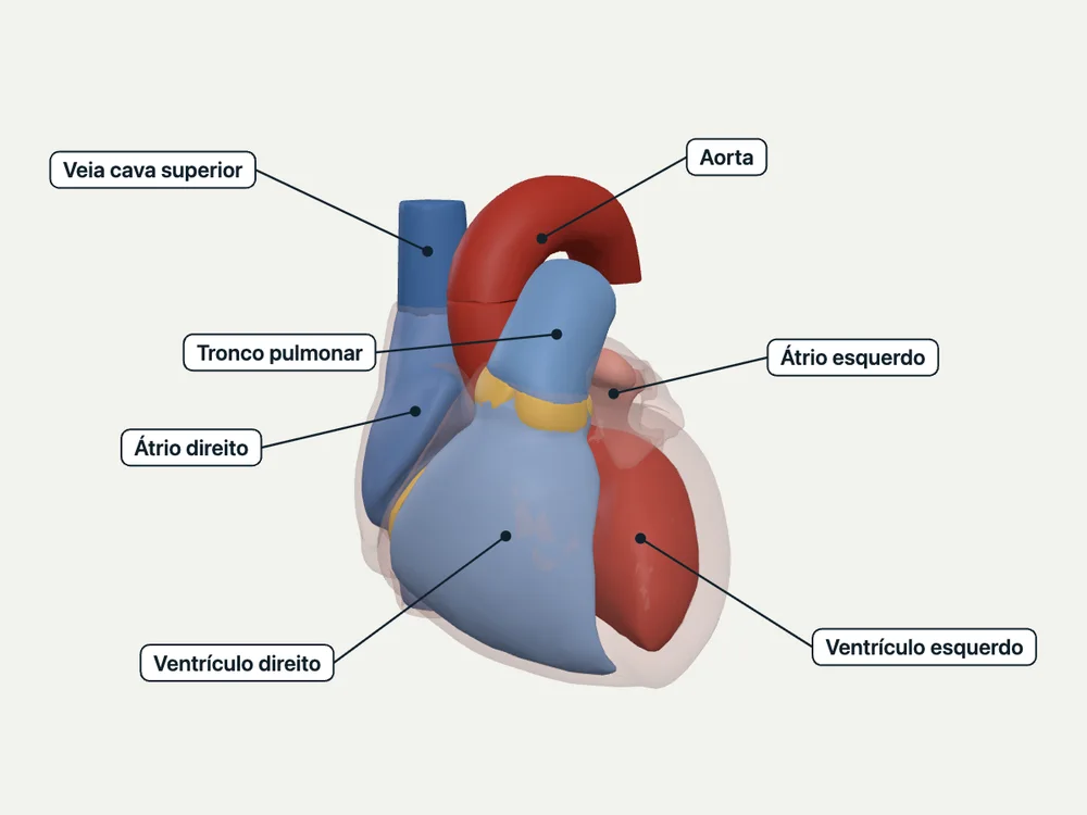
O coração são duas bombas em série — a direita empurrando contra a resistência do circuito pulmonar, a esquerda contra a do sistêmico —, e a forma de cada peça segue essa função: cordas tendíneas e músculos papilares travam as valvas atrioventriculares no instante da sístole, a fossa ovalis é o ponto mais fino do septo interatrial, o septo interventricular é a única via elétrica entre átrios e ventrículos, e as valvas semilunares nascem de uma linhagem embrionária própria (o truncus arteriosus), não da parede ventricular. Esta unidade percorre essas cinco peças com fonte, e declara por escrito onde a fonte para e a inferência começa.
Educational material written with AI support and checked by automated review, without a named medical reviewer. It does not replace the textbook or clinical guidance.
Essential
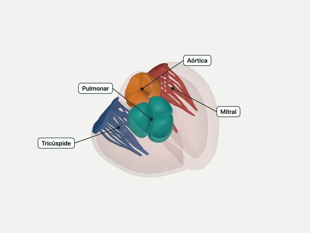
As cúspides das valvas atrioventriculares (tricúspide, à direita; mitral, à esquerda) recebem cordas tendíneas finas que as prendem a músculos papilares nascidos da própria parede do ventrículo correspondente — 'they arise from the inner walls of the left and right ventricle and attach to mitral and tricuspid valve leaflets respectively via chordae tendinae'.
Quando o ventrículo contrai na sístole, os músculos papilares contraem JUNTO — não antes, não depois — e essa contração simultânea tensiona as cordas: 'when the ventricles contract during the systole, simultaneous contraction of the papillary muscles taut the chordae tendinae. This arrangement prevents prolapse as well as the inversion of the cusps of the mitral and tricuspid valves.' A corda tensionada segura a cúspide no lugar bem no momento em que a pressão ventricular está mais alta e mais empurraria a cúspide de volta para o átrio — é um freio mecânico sincronizado com a própria contração que ele existe para conter.
A fossa ovalis fica na porção inferior do septo interatrial, do lado do átrio direito, e é a única parte desse septo que forma o chamado 'septo verdadeiro' — apenas 20% da área septal inteira: 'the true interatrial septum contains the fossa ovalis and comprises only 20% of the entire septum. It is the only area through which the interatrial septum may be traversed without the risk of cardiac perforation.'
Ser a parte mais fina E a única atravessável sem risco de perfuração é exatamente o que faz da fossa ovalis o alvo de escolha para quem precisa atravessar o septo de propósito: 'the (limbus) fossa ovalis represents the most direct anatomical landmark for atrial septal puncture for various procedures' — cateterismo transeptal, valvuloplastia por balão, ablação por radiofrequência, isolamento de veias pulmonares. Nenhuma outra parte do septo oferece essa combinação (fina + segura), e é por isso que o restante do septo NÃO é usado para esses procedimentos.
O miocárdio é mais espesso nos ventrículos do que nos átrios — 'though it is thicker in the ventricles and thinner in the atria. This disparity is due to the differences in the generated force of contraction needed for propelling blood between the atria and the ventricles, with the ventricles requiring much more power' — e a espessura da parede aumenta quando a câmara passa a enfrentar mais pressão para vencer: 'chronically high afterloads from higher mean arterial pressures cause the left ventricular muscle to contract against greater pressures, leading to compensatory cardiomyocyte hypertrophy. The ventricular muscle wall thickens to reduce wall stress.'
DECLARADO: nenhuma das cinco fontes desta unidade compara, em número ou em frase direta, a espessura BASAL do ventrículo esquerdo com a do direito — a citação acima descreve hipertrofia ventricular esquerda PATOLÓGICA por hipertensão crônica, não a diferença normal entre os dois ventrículos. O que se deriva daqui é o PRINCÍPIO, não o número: parede mais espessa é resposta a mais pressão a vencer. Aplicando esse mesmo princípio à distinção que a visão geral do coração estabelece — o ventrículo direito empurra sangue para o circuito PULMONAR (via valva pulmonar e tronco pulmonar) e o esquerdo para o circuito SISTÊMICO (via valva aórtica e aorta) —, a expectativa é que a câmara que enfrenta a maior resistência crônica (o ventrículo esquerdo, contra a circulação sistêmica) tenha, também em condições basais, a parede mais espessa. É uma inferência a partir do princípio citado, não uma afirmação que alguma das cinco fontes faça por extenso — e fica marcada como tal.
O septo interventricular não é só a parede que separa os dois ventrículos — é também a única passagem elétrica entre átrios e ventrículos. Depois de cruzar o nó atrioventricular, o impulso entra nele: 'the AV bundles carrying impulses from the AV node cross the fibrous ring to enter the interventricular septum, and divides into the right and left bundles of His.'
Cada um dos dois ramos percorre o septo até a base dos músculos papilares correspondentes antes de se ramificar nas fibras de Purkinje: 'right bundle of His supply, the musculature of the right ventricle, and the left bundle supply the left ventricle. Each of these bundles further divides into thin fibers called Purkinje fibers at the base of papillary muscles.' Fora dessa via, o anel fibroso que separa átrios de ventrículos é eletricamente isolante — então o septo interventricular é o único corredor por onde o impulso passa de um lado do coração ao outro.
In the clinic
Isquemia, fibrose pós-infarto ou ruptura do músculo papilar tiram esse freio: 'any morphological changes in these muscles and loss of contractility like in myocardial infarction with muscle fibrosis or ischemia can result in a malfunction of these valves leading to regurgitation'. Sem o músculo papilar contraindo em sincronia, a cúspide perde o ponto de ancoragem tensionado e prolapsa para o átrio na sístole — regurgitação mitral ou tricúspide aguda, e a fonte é direta sobre a gravidade quando o músculo rompe de vez: 'the rupture of papillary muscles should receive immediate treatment; not doing so can be fatal'.
A mesma fragilidade embrionária que faz a fossa ovalis existir (fusão do septo primum com o septo secundum) é o que pode falhar e deixar um forame oval patente — canal residual entre os dois átrios. Clinicamente isso aparece como substrato de embolia paradoxal: 'most patients with cryptogenic stroke have a patent foramen ovale which serves as a channel for paradoxical emboli leading to a greater incidence of cerebrovascular accidents.'
Quando a pós-carga do ventrículo esquerdo sobe de forma crônica (hipertensão arterial sistêmica sustentada), a parede espessa ainda mais — hipertrofia ventricular esquerda concêntrica —, e isso tem achado de exame físico descrito na própria fonte: 'physical examination findings of left ventricular hypertrophy may include a diffuse point of maximal impulse and an S4 gallop upon cardiac apex auscultation.'
A porção membranosa do septo interventricular — a mesma região atravessada pelo feixe AV — é o local mais comum de comunicação interventricular congênita, isolada ou como um dos quatro achados da tetralogia de Fallot: 'tetralogy of Fallot consists of a combination of VSD of the membranous portion of the interventricular septum, stenosis of the orifice of the pulmonary artery, the aortic orifice overriding the VSD, and hypertrophy of the right ventricle.'
Open the deeper layer
Going deeper
O truncus arteriosus (via de saída embrionária única do coração primitivo) e as cristas conotruncais que crescem em espiral dentro dele: 'the formation of the aorta and the pulmonary trunk is dependent upon the truncus arteriosus. During development, the right and left conotruncal ridges grow in a spiral, twisted fashion forming the great vessels.'
Aorta ascendente — metade do truncus separada pelo septo aorticopulmonar: 'an aorticopulmonary septum will result in the separation of the Aorta and pulmonary trunk.'; Tronco pulmonar — a outra metade da mesma separação, continuação da via de saída do ventrículo direito.; Valva aórtica (semilunar) — formada pelas células mesenquimais da mesma crista conotruncal: 'this formation of the ridges is under the guidance of the combination of the mesenchymal cells that are going to create the semilunar valves (aortic and pulmonary semilunar valves).'; Valva pulmonar (semilunar) — formada pelo mesmo processo, do lado pulmonar da separação.
Test yourself before moving on
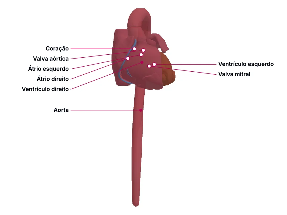
Uma hemácia entra no coração pela veia cava superior. Nomeie, na ordem, cada câmara e cada valva que ela atravessa até sair pela aorta.
The answer stays in Assessment, with no spoiler here.
Answer in AssessmentSources (5)
- Rich NL, Khan YS. Anatomy, Thorax, Heart Papillary Muscles. [Updated 2023 Jul 24]. In: StatPearls [Internet]. Treasure Island (FL): StatPearls Publishing; 2026 Jan-.
They arise from the inner walls of the left and right ventricle and attach to mitral and tricuspid valve leaflets respectively via chordae tendinae.
- Oduah MTA, Sharma P, Brown KN. Anatomy, Thorax, Heart Fossa Ovalis. [Updated 2023 Jul 25]. In: StatPearls [Internet]. Treasure Island (FL): StatPearls Publishing; 2026 Jan-.
The fossa ovalis is a depressed structure located in the lower portion of the right atrial side of the interatrial septum.
- Tran DB, Weber C, Lopez RA. Anatomy, Thorax, Heart Muscles. [Updated 2024 Jun 22]. In: StatPearls [Internet]. Treasure Island (FL): StatPearls Publishing; 2026 Jan-.
Myocardium is found in the walls of all 4 heart chambers, though it is thicker in the ventricles and thinner in the atria. This disparity is due to the differences in the generated force of contraction needed for propelling blood between the atria and the ventricles, with the ventricles requiring mu…
- Rehman I, Rehman A. Anatomy, Thorax, Heart. [Updated 2023 Aug 28]. In: StatPearls [Internet]. Treasure Island (FL): StatPearls Publishing; 2026 Jan-.
The right ventricle pumps blood through the right ventricular outflow tract, across the pulmonic valve, and into the pulmonary artery that distributes it to the lungs for oxygenation.
- Bamalan OA, Jozsa F, Soos MP. Anatomy, Thorax, Heart Great Vessels. [Updated 2023 Jul 25]. In: StatPearls [Internet]. Treasure Island (FL): StatPearls Publishing; 2026 Jan-.
The formation of the aorta and the pulmonary trunk is dependent upon the truncus arteriosus. During development, the right and left conotruncal ridges grow in a spiral, twisted fashion forming the great vessels.
More Anatomia summaries
After reading, use what you learned
Atlas
See the structures of the topic in 3D and move through the layers.
Clinical case
Apply the content to an on-call decision.
Exam
Read the result that closes the reasoning.
Assessment
Answer the block questions and see where you stand.
AI tutor
Ask about what the text did not settle.
Start free: create an account and try a sample of the 3D Atlas, the clinical case, the exam, the assessment and the AI tutor. No credit card.
Start free