O membro superior, do ombro à mão
O manguito rotador - subescapular, infraespinal, redondo menor e supraespinal (SITS) - centraliza uma cabeça umeral grande demais para a cavidade glenoide RASA da escápula, trocando estabilidade óssea por mobilidade: por isso o ombro é a articulação mais luxada do corpo (direção anteroinferior em mais de 95% dos casos), o que arrisca o nervo axilar no espaço quadrangular (borda pelo redondo menor), testável pela sensibilidade do 'distintivo regimental'.
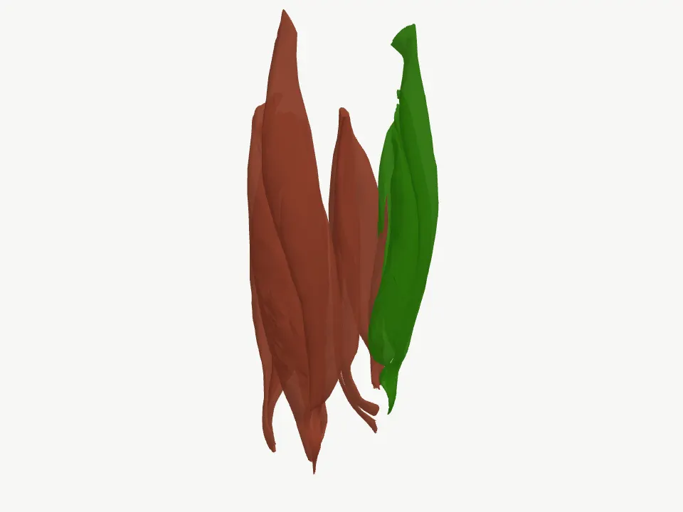
Ombro: cíngulo escapular, úmero e manguito rotador
O manguito rotador - subescapular, infraespinal, redondo menor e supraespinal (SITS) - centraliza uma cabeça umeral grande demais para a cavidade glenoide RASA da escápula, trocando estabilidade óssea por mobilidade: por isso o ombro é a articulação mais luxada do corpo (direção anteroinferior em mais de 95% dos casos), o que arrisca o nervo axilar no espaço quadrangular (borda pelo redondo menor), testável pela sensibilidade do 'distintivo regimental'. O supraespinal, que inicia a abdução, atravessa um espaço subacromial estreito e está entre os tendões mais comumente lesados do manguito, com fraqueza testável pelo teste de Jobe. A clavícula, elo do cíngulo peitoral com a escápula, está entre os ossos mais comumente fraturados do corpo, tipicamente no terço médio - seu segmento mais fino e sem reforço ligamentar.
What it is. A cavidade glenoide - parte da própria malha da escápula, não uma peça à parte - é rasa: 'The glenoid cavity is a shallow osseous structure deepened by a fibrocartilaginous rim, the glenoid labrum, which spans the vault's osseous periphery.' O tamanho da cabeça umeral em relação a essa cavidade rasa é desproporcional: 'The humeral head's disproportionate size relative to the shallow glenoid fossa (a 4:1 surface area ratio) contribute to the shoulder joint's high mobility and susceptibility to dislocation.'
Why it matters. Quem compensa essa falta de encaixe ósseo são os quatro músculos do manguito rotador - subescapular, infraespinal, redondo menor e supraespinal, o mnemônico SITS: 'The rotator cuff includes the following muscles: Subscapularis Infraspinatus Teres minor Supraspinatus A helpful mnemonic for recalling these muscles is "SITS."' Eles se originam cada um de uma região distinta da MESMA escápula e convergem sobre o úmero: 'The primary biomechanical role of the rotator cuff is to stabilize the glenohumeral joint through compression of the humeral head against the glenoid. The 4 rotator cuff muscles originate from the scapula and insert on the humerus.' Coordenados, eles mantêm a cabeça umeral centralizada durante o movimento: 'Coordinated contraction of the rotator cuff muscles maintains humeral head centering within the glenoid during shoulder motion, permitting a broad range of motion while preserving joint stability.' - e é por isso que a fonte os descreve como 'The rotator cuff muscles thus act as the glenohumeral joint's dynamic stabilizers.' Mas essa cobertura muscular não é completa: 'leaving the inferior aspect relatively unsupported. This anatomic configuration increases the risk of inferior glenohumeral dislocation.'
What it is. Dos quatro músculos do manguito, o supraespinal é quem inicia o movimento de abdução: 'The supraspinatus muscle originates in the supraspinous fossa of the scapula and inserts into the greater tubercle of the humerus, facilitating the abduction of the humerus.' e 'The supraspinatus initiates shoulder abduction and is particularly important during the first 0° to 15° of motion, after which the deltoid becomes the principal abductor.'
Why it matters. Para chegar ao tubérculo maior, o tendão do supraespinal atravessa um corredor estreito sob o acrômio: 'The subacromial space is bounded superiorly by the acromion and acromioclavicular joint, anteriorly by the acromion and coracoacromial ligament, and inferiorly by the humeral head. Within this space lie the supraspinatus tendon, portions of the infraspinatus and subscapularis tendons, the long head of the biceps tendon, and the subacromial-subdeltoid bursa.' Esse espaço já é apertado por natureza: 'The subacromial space, typically 1 to 1.5 cm in height, narrows physiologically with abduction and forward flexion.' E o próprio mecanismo que deveria protegê-lo pode piorar o aperto: quando o manguito enfraquece ou se fatiga, a cabeça umeral perde a força que a mantém centralizada e migra para cima, estreitando ainda mais o espaço que o supraespinal precisa atravessar: 'Secondary impingement occurs despite normal static anatomy and results from dynamic factors that allow superior migration of the humeral head during motion, such as rotator cuff weakness or fatigue resulting in loss of centralizing force'
What it is. A direção anterior domina as luxações de ombro: 'Anterior dislocation comprises 96% of cases and is often the result of a force directed to the shoulder joint while the arm is abducted and externally rotated.' - confirmado de outra fonte: 'Over 95% of shoulder instability events occur in the anterior direction.' O nervo axilar contorna exatamente a região que essa luxação estica: 'The axillary nerve, formed from the posterior cord of the brachial plexus, wraps around the surgical neck of the humerus, and provides innervation to the deltoid and rotator cuff muscles, specifically the teres minor.' - 'The axillary nerve, which wraps around the humeral surgical neck, can sustain damage during dislocation or reduction attempts.'
Why it matters. Antes de alcançar o deltoide, o nervo axilar passa por um espaço quadrangular estreito, cuja borda superior é o próprio redondo menor: 'The quadrangular space is bordered superiorly by the lower border of teres minor muscle, laterally by the surgical neck of the humerus, inferiorly by the upper boundary of teres major, and medially by the long head of the triceps brachial.' É justamente nesse trajeto - e na cápsula anteroinferior que a luxação distende - que o nervo é mais vulnerável: 'The axillary nerve is most susceptible to injury at the origin from the posterior cord, in the quadrilateral space, the anteroinferior aspect of the shoulder capsule, and within the subfascial surface of the deltoid muscle.' Por isso ele é 'The axillary nerve is the most common damaged nerve in glenohumeral dislocation' e, cirurgicamente, 'The axillary nerve (most common nerve injury) is adjacent to the inferior capsule and closest to the inferior glenoid rim at the 6 o'clock position.'
What it is. A clavícula fecha o cíngulo peitoral pela frente: 'The clavicle is a sigmoid-shaped long bone with a convex surface along its medial end when observed from a cephalad position. The clavicle connects the axial and appendicular skeletons and, along with the scapula, forms the pectoral girdle.' E, entre todos os ossos do corpo, 'The clavicle is one of the most commonly fractured bones in the human body.'
Why it matters. A razão é geométrica, não só de exposição: 'Because the midshaft of the clavicle is the thinnest segment and does not contain ligamentous attachments, it is the most easily fractured aspect of the clavicle.' Esse ponto mais fino - sem o reforço ligamentar que protege as extremidades - concentra a maioria das fraturas: 'The middle third of the clavicle is fractured in 69% of cases, the distal third is fractured in 28% of cases, and the proximal third is fractured in 3% of cases.'
When it fails. Estabilidade dependente de músculo, e não de osso, tem um preço: 'The glenohumeral joint is by far the most frequently dislocated joint of the body, accounting for up to 45% of dislocations.' e, quando ocorre, 'Anterior dislocation comprises 96% of cases and is often the result of a force directed to the shoulder joint while the arm is abducted and externally rotated.'
When it fails. Não por acaso, 'In adults, rotator cuff injury is the most common tendon injury seen and treated.' A fraqueza resultante é testável à beira do leito: 'The Jobe or empty can test used to evaluate for supraspinatus tendinopathy. The patient elevates the arm to about 90 degrees, and with the thumb down (having an empty can in the patient's hand), isometric force is applied downward.' - descrito de outra fonte como 'With the arms in 90° of abduction in the scapular plane and internally rotated (thumbs down), the patient resists downward pressure. Pain and/or weakness suggest supraspinatus pathology.'
When it fails. A lesão desse nervo tem sinal sensitivo específico e testável: 'The upper lateral cutaneous nerve provides sensation to the inferior lateral deltoid region, at an area called the regimental badge area.' - achado que integra o quadro descrito como 'Axillary nerve injury may lead to deltoid paralysis and loss of sensation over the lateral shoulder.' No plano motor, é o próprio redondo menor - cuja borda forma o espaço por onde o nervo passa - que ajuda a manter a cabeça umeral na cápsula: 'Teres minor is one of the muscles which stabilize the glenohumeral joint. It acts by laterally rotating the arm and helps to keep the head of the humerus in the shoulder capsule.'
When it fails. O mecanismo típico reforça o padrão: 'These injuries most often result from a fall onto the lateral shoulder, which transmits force along the length of the bone.'
+ Going deeper
Where it comes from. A escápula tem uma concavidade na face ventral, a fossa subescapular: 'The ventral surface of the scapula abuts the thoracic rib cage and has a large concavity called the subscapular fossa where the subscapularis attaches.' Na face dorsal, a espinha da escápula separa duas outras fossas: 'Above the spine is the supraspinous fossa and below it is the infraspinous fossa.' E uma quarta origem fica fora das fossas, na borda lateral: 'A ridge runs down the infraspinous fossa near the lateral border from the glenoid cavity to just above the inferior angle. This ridge separates the teres major and minor muscles from the infraspinatus muscle.'
What comes with it. Subescapular - origina-se na fossa subescapular (face ventral) e se insere no tubérculo menor do úmero: 'The muscle originates from the subscapular fossa and inserts onto the lesser tubercle of the humerus.' É o único rotator interno do grupo SITS: 'The subscapularis is the primary medial (internal) rotator of the shoulder, whereas the infraspinatus and teres minor function primarily as lateral (external) rotators.' e o maior componente da parede posterior da axila, prevenindo a luxação anterior durante a abdução: 'The subscapularis is the largest component of the posterior wall of the axilla. This muscle prevents anterior dislocation of the humerus during abduction and produces medial rotation of the arm.'; Supraespinal - origina-se na fossa supraespinal e se insere no tubérculo maior, iniciando a abdução do ombro: 'The supraspinatus muscle originates in the supraspinous fossa of the scapula and inserts into the greater tubercle of the humerus, facilitating the abduction of the humerus.' - 'The supraspinatus initiates shoulder abduction and is particularly important during the first 0° to 15° of motion, after which the deltoid becomes the principal abductor.'; Infraespinal - origina-se na fossa infraespinal e na espinha da escápula, inserindo-se no tubérculo maior e permitindo rotação externa: 'The infraspinatus muscle originates in the infraspinous fossa and scapula spine and inserts into the greater tubercle of the humerus as well and allows external rotation of the humerus.'; Redondo menor - origina-se na borda lateral da escápula, fora das três fossas, e se insere no tubérculo maior, permitindo rotação externa: 'The teres minor originates on the lateral border of the scapula and inserts into the greater tubercle and allows external rotation.' Sua borda superior fecha o espaço quadrangular por onde passam o nervo axilar e os vasos circunflexos posteriores do úmero: 'The quadrangular space is bordered superiorly by the lower border of teres minor muscle, laterally by the surgical neck of the humerus, inferiorly by the upper boundary of teres major, and medially by the long head of the triceps brachial.' e o músculo ajuda a manter a cabeça umeral na cápsula: 'Teres minor is one of the muscles which stabilize the glenohumeral joint. It acts by laterally rotating the arm and helps to keep the head of the humerus in the shoulder capsule.'
Um homem de 68 anos relata dor crônica no ombro e dificuldade progressiva para levantar o braço para pentear o cabelo. No exame, com o braço a 90° de abdução e o polegar para baixo, ele não consegue resistir a uma pressão leve para baixo. Qual músculo esse teste isola, e por que ele - entre os quatro do manguito - é o mais vulnerável a esse tipo de lesão crônica?
Answer in AssessmentThe answer stays in Assessment, with no spoiler here.Sources for this block (11)
- Chang LR, Anand P, Varacallo MA. Anatomy, Shoulder and Upper Limb, Glenohumeral Joint. [Updated 2025 Mar 3]. In: StatPearls [Internet]. Treasure Island (FL): StatPearls Publishing; 2026 Jan-.
“The glenoid cavity is a shallow osseous structure deepened by a fibrocartilaginous rim, the glenoid labrum, which spans the vault's osseous periphery.”
- La Rose J, Madrazo-Ibarra A, Varacallo MA. Anatomy, Rotator Cuff. [Updated 2026 Jun 18]. In: StatPearls [Internet]. Treasure Island (FL): StatPearls Publishing; 2026 Jan-.
“The rotator cuff includes the following muscles: Subscapularis Infraspinatus Teres minor Supraspinatus A helpful mnemonic for recalling these muscles is "SITS."”
- Mostafa E, Imonugo O, Varacallo MA. Anatomy, Shoulder and Upper Limb, Humerus. [Updated 2023 Aug 7]. In: StatPearls [Internet]. Treasure Island (FL): StatPearls Publishing; 2026 Jan-.
“The supraspinatus muscle originates in the supraspinous fossa of the scapula and inserts into the greater tubercle of the humerus, facilitating the abduction of the humerus.”
- Creech JA, Busse A, Li D, Pinkerman S, Silver S. Shoulder Impingement Syndrome. [Updated 2026 Jan 10]. In: StatPearls [Internet]. Treasure Island (FL): StatPearls Publishing; 2026 Jan-.
“The subacromial space is bounded superiorly by the acromion and acromioclavicular joint, anteriorly by the acromion and coracoacromial ligament, and inferiorly by the humeral head. Within this space lie the supraspinatus tendon, portions of the infraspinatus and subscapularis tendons, the long head …”
- May T, Garmel GM. Rotator Cuff Injury. [Updated 2023 Jun 26]. In: StatPearls [Internet]. Treasure Island (FL): StatPearls Publishing; 2026 Jan-.
“In adults, rotator cuff injury is the most common tendon injury seen and treated.”
- Varacallo MA, Musto MA, Mair SD. Anterior Shoulder Instability. [Updated 2023 Aug 4]. In: StatPearls [Internet]. Treasure Island (FL): StatPearls Publishing; 2026 Jan-.
“Over 95% of shoulder instability events occur in the anterior direction.”
- Okwumabua E, Thompson JH. Anatomy, Shoulder and Upper Limb, Axillary Nerve. [Updated 2023 Jul 24]. In: StatPearls [Internet]. Treasure Island (FL): StatPearls Publishing; 2026 Jan-.
“The quadrangular space is bordered superiorly by the lower border of teres minor muscle, laterally by the surgical neck of the humerus, inferiorly by the upper boundary of teres major, and medially by the long head of the triceps brachial.”
- Tessler J, Talati R. Axillary Nerve Injury. In: StatPearls [Internet]. Treasure Island (FL): StatPearls Publishing; 2026 Jan-.
“The axillary nerve is most susceptible to injury at the origin from the posterior cord, in the quadrilateral space, the anteroinferior aspect of the shoulder capsule, and within the subfascial surface of the deltoid muscle.”
- La Rose J, Charlick M, Varacallo MA. Anatomy, Shoulder and Upper Limb, Clavicle. [Updated 2026 Jul 15]. In: StatPearls [Internet]. Treasure Island (FL): StatPearls Publishing; 2026 Jan-.
“The clavicle is a sigmoid-shaped long bone with a convex surface along its medial end when observed from a cephalad position. The clavicle connects the axial and appendicular skeletons and, along with the scapula, forms the pectoral girdle.”
- Le MQT, Myers K, May T, Hosseinzadeh S. Clavicle Fracture. [Updated 2026 Mar 27]. In: StatPearls [Internet]. Treasure Island (FL): StatPearls Publishing; 2026 Jan-.
“Because the midshaft of the clavicle is the thinnest segment and does not contain ligamentous attachments, it is the most easily fractured aspect of the clavicle.”
- Miniato MA, Mudreac A, Borger J. Anatomy, Thorax, Scapula. [Updated 2023 Jul 24]. In: StatPearls [Internet]. Treasure Island (FL): StatPearls Publishing; 2026 Jan-.
“The ventral surface of the scapula abuts the thoracic rib cage and has a large concavity called the subscapular fossa where the subscapularis attaches.”
Escápula alada: dois padrões de winging e por que se confundem
A escápula fica presa à parede torácica pelo equilíbrio de três músculos que puxam em direções diferentes - serrátil anterior (protrai), trapézio (eleva/retrai/deprime) e romboides (retraem e fixam a borda medial) - e perder qualquer um deles rompe esse equilíbrio, produzindo escápula alada ('winging'). O padrão MEDIAL (escápula translada para cima, ângulo inferior gira em direção à coluna) vem da paralisia do serrátil anterior por lesão do nervo torácico longo, cujo trajeto longo e superficial na parede lateral do tórax o expõe tanto à dissecção axilar/mastectomia quanto a esportes de arremesso - é a causa mais comum de todas. O padrão LATERAL (escápula translada para baixo, ângulo inferior gira para longe da coluna) vem classicamente da paralisia do trapézio por lesão do nervo acessório espinhal no triângulo posterior do pescoço (biópsia de linfonodo cervical é a causa iatrogênica clássica), mas também pode vir dos romboides (nervo dorsal da escápula, raiz C5 isolada) - uma causa mais rara e um achado tipicamente sutil, subdiagnosticado pela posição profunda do músculo.
What it is. O serrátil anterior se origina nas primeiras costelas e se insere na escápula: 'The serratus anterior is a fan-shaped muscle that originates on the superolateral surfaces of the first to eighth ribs or the first to ninth ribs at the lateral wall of the thorax and inserts along the superior angle, medial border, and inferior angle of the scapula.' Sua ação é puxar a escápula para frente ao redor do tórax: 'The serratus anterior muscle pulls the scapula forward around the thorax, which allows for anteversion and protraction of the arm'
Why it matters. Quem inerva esse músculo é o nervo torácico longo, formado pelas raízes C5 a C7: 'The long thoracic nerve, which originates from the upper portion of the superior trunk of the brachial plexus and is typically composed of cervical nerves C5, C6, and C7, is responsible for the innervation of the serratus anterior.' Esse nervo corre de forma longa e superficial sobre a própria superfície do músculo que inerva: 'The long thoracic nerve courses inferiorly on the surface of the serratus anterior and, as such, is susceptible to damage during specific surgical procedures, especially with axillary lymph node dissection.' e 'The long thoracic nerve is at risk of compression due to its superficial course along the lateral chest wall.' - uma exposição que o torna vulnerável tanto a procedimentos cirúrgicos quanto a esportes: 'Thoracic procedures, such as radical mastectomy, transthoracic sympathectomy, misplaced intercostal drains, axillary lymph node dissections, and resection of the first rib, increase the exposure of the long thoracic nerve and risk of nerve injury.' e 'The sports involved in serratus anterior paralysis include archery, baseball, basketball, bodybuilding, bowling, football, golf, gymnastics, hockey, soccer, tennis, and wrestling.'
What it is. O trapézio tem três porções funcionais distintas. A superior eleva e roda a escápula para cima: 'The upper fibers can elevate and upwardly rotate the scapula and extend the neck.' A média aduz as duas escápulas: 'The middle portion attaches to the upper part of the spinous processes of C7-T12. This portion adducts (draws together) the 2 scapulae.' E a inferior deprime a escápula e ajuda a rotação para cima: 'The lower fibers depress and assist the upper fibers in the upward rotation of the scapula.'
Why it matters. A função motora das três porções depende de um único nervo: 'The ventral rami of C3 and C4 innervate the sensory functions of the trapezius. The spinal accessory nerve (cranial nerve XI) innervates the motor function of the trapezius.' Esse nervo atravessa uma região superficial e estreita do pescoço antes de alcançar o músculo: 'The spinal accessory nerve then passes through the posterior triangle of the neck.' - 'The 3 sides of the triangle are formed by the sternocleidomastoid, trapezius, and clavicle. The nerve then passes beneath the trapezius, where it innervates it.' Uma lesão que ocorra justamente dentro dessa região tem uma consequência precisa: 'Only the trapezius is denervated if the nerve is injured within the posterior triangle.' É exatamente esse cenário que torna certos procedimentos cirúrgicos a causa iatrogênica mais comum de paralisia do trapézio: 'Iatrogenic injury to the spinal accessory nerve during cervical lymph node biopsy or excision is the most common cause of trapezius palsy.'
What it is. Os romboides são dois músculos - maior e menor - com origem e inserção próprias. O menor: 'The rhomboid minor is a cylindrical muscle that originates at the ligamentum nuchae and C7 and T1 vertebra. It inserts at the scapula's medial border near the base of the spine of the scapula.' O maior: 'The rhomboid major is a quadrangular muscle located inferior to the rhomboid minor. The origin of the rhomboid muscles is from the spinous processes of the T2-T5 vertebra and inserts on the medial border of the scapula, just inferior to the rhomboid minor.' Juntos, sua ação é: 'Functionally, the rhomboid muscles retract, elevate and rotate the scapula. They also protract the medial border of the scapula, keeping it in position at the posterior thoracic wall.'
Why it matters. A inervação motora dos dois vem de um único nervo: 'The motor function of the rhomboid muscles is controlled by the dorsal scapular nerve (DSN). The dorsal scapular nerve originates from the ventral ramus of the spinal nerve root C5 and courses posterior inferiorly through the middle scalene muscles and between the posterior scalene, levator scapulae (to which it also provides innervation), and the serratus posterior superior.' Esse nervo nasce isoladamente de UMA raiz cervical - sem passar pelos troncos do plexo braquial, como acontece com o nervo torácico longo (C5-C7) ou o acessório espinhal (que desce do crânio): 'The dorsal scapular nerve (DSN) is a motor nerve arising from the ventral ramus of the C5 nerve root. Once branching from the brachial plexus, the DSN pierces the middle scalene muscle, coursing deep to the levator scapulae and the rhomboid major and minor muscles.' e 'The dorsal scapular nerve is a motor nerve that innervates the levator scapulae, rhomboid major, and rhomboid minor muscles.'
What it is. Os três músculos - serrátil anterior, trapézio e romboides - cumprem a mesma função geral, cada um a seu modo: 'These muscles contribute to maintaining the scapula protracted against the posterior thoracic wall. Neurologic insult to any of these muscles results in winging of the scapula as it lifts off the thoracic wall.' É por isso que a maioria dos casos de escápula alada nasce de apenas dois deles: 'Most cases are secondary to neurologic lesions involving the serratus anterior and trapezius muscles. Scapular winging may be caused less frequently by neurologic lesions involving the rhomboid muscles.'
Why it matters. A aparência geral - a escápula se soltando da parede torácica - é a mesma nos dois padrões, mas a DIREÇÃO do deslocamento diferencia um do outro com precisão: 'Long thoracic nerve injury results in serratus anterior muscle paralysis and causes medial scapular winging. The scapula translates superiorly, and the inferior angle rotates medially. Spinal accessory nerve injury results in trapezius muscle paralysis and causes lateral scapular winging. The scapula translates inferiorly, and the inferior angle rotates laterally.' A explicação fisiológica para essa diferença de direção está no equilíbrio de forças entre os três músculos: 'A weakness of the serratus anterior muscle is the most prevalent cause of winging due to the weak protraction of the scapula and unopposed medial scapula retraction by the rhomboid muscles and elevation by the trapezius, resulting in medial winging of the scapula. This condition is easily differentiated from the much less common lateral winging due to weakness of the trapezius muscle, which is supplied by the spinal accessory nerve.' Resumindo a distinção entre os padrões: 'Medial scapular winging typically results from weakness of the serratus anterior muscle, often caused by injury to the long thoracic nerve. In contrast, lateral winging arises from dysfunction of the trapezius or rhomboid muscles, associated with spinal accessory or dorsal scapular nerve impairment.'
When it fails. Quando o nervo torácico longo é lesado, o serrátil anterior não consegue mais segurar a escápula contra a caixa torácica: 'If the long thoracic nerve is damaged, a clinical phenomenon known as a winged scapula occurs as the serratus anterior is no longer able to hold the scapula against the chest wall. The winging is medial scapula winging that has to be differentiated from lateral winging due to a weakness of the rhomboids or the trapezius muscles.' Esse é, de fato, o padrão mais comum de todos: 'The most common cause of scapular winging is serratus anterior paralysis after an insult to the long thoracic nerve.'
When it fails. A paralisia do trapézio por lesão do nervo acessório espinhal produz um padrão de winging bem definido: 'Lateral scapular winging results from a deficit in trapezius muscle function secondary to injury to the spinal accessory nerve.' - com uma direção específica de deslocamento: 'Long thoracic nerve injury results in serratus anterior muscle paralysis and causes medial scapular winging. The scapula translates superiorly, and the inferior angle rotates medially. Spinal accessory nerve injury results in trapezius muscle paralysis and causes lateral scapular winging. The scapula translates inferiorly, and the inferior angle rotates laterally.'
When it fails. A paralisia dos romboides também entra na categoria de winging lateral, mas com uma frequência e uma clareza clínica bem diferentes das do trapézio: 'Lateral scapular winging can also result from a deficit in rhomboid muscle function secondary to injury to the dorsal scapular nerve. Rhomboid paralysis is less common than trapezius paralysis.' Epidemiologicamente, 'Rhomboid paralysis is also rare, and its associated functional impairments are even less common.' E quando o achado aparece, ele costuma ser difícil de reconhecer: 'Physical examination often yields subtle lateral winging of the scapula. As a result of the subtle exam finding and the deep anatomic position of the rhomboids, rhomboid palsy is often underdiagnosed.'
When it fails. Como a inspeção visual sozinha pode ser ambígua, a diferenciação clínica usa testes de força que isolam cada músculo especificamente: 'Trapezius muscle strength can be tested with resisted elevation of the scapula. The function of the rhomboid and levator scapulae is evaluated by having the patient place both hands on the hips and pushing the elbows posteriorly against resistance. Serratus anterior strength can be assessed with the forward shoulder flexion to 90° while the patient performs push-ups.'
+ Going deeper
Where it comes from. A escápula não fica presa à caixa torácica por encaixe ósseo - ela é mantida ali por músculos que a protraem contra a parede torácica posterior: 'These muscles contribute to maintaining the scapula protracted against the posterior thoracic wall. Neurologic insult to any of these muscles results in winging of the scapula as it lifts off the thoracic wall.' Qualquer um desses músculos, ao falhar, permite que a escápula se solte: 'Most cases are secondary to neurologic lesions involving the serratus anterior and trapezius muscles. Scapular winging may be caused less frequently by neurologic lesions involving the rhomboid muscles.'
What comes with it. Serrátil anterior - inervado pelo nervo torácico longo (C5-C7), de curso longo e superficial ao longo da parede lateral do tórax: 'The long thoracic nerve courses inferiorly on the surface of the serratus anterior and, as such, is susceptible to damage during specific surgical procedures, especially with axillary lymph node dissection.' Sua paralisia é a causa mais comum de escápula alada, com padrão MEDIAL: 'The most common cause of scapular winging is serratus anterior paralysis after an insult to the long thoracic nerve.'; Trapézio - inervado pelo nervo acessório espinhal (com contribuição de C3-C4 do plexo cervical), que atravessa o triângulo posterior do pescoço antes de alcançar o músculo, e cuja lesão ali denerva SÓ o trapézio: 'Only the trapezius is denervated if the nerve is injured within the posterior triangle.' A biópsia de linfonodo cervical nesse ponto é a causa iatrogênica mais comum de paralisia isolada do trapézio, com padrão LATERAL: 'Iatrogenic injury to the spinal accessory nerve during cervical lymph node biopsy or excision is the most common cause of trapezius palsy.'; Romboides (maior e menor) - inervados por um único nervo, o dorsal da escápula, que nasce isoladamente da raiz ventral de C5 e corre profundo, sob o levantador da escápula e os próprios romboides: 'The dorsal scapular nerve (DSN) is a motor nerve arising from the ventral ramus of the C5 nerve root. Once branching from the brachial plexus, the DSN pierces the middle scalene muscle, coursing deep to the levator scapulae and the rhomboid major and minor muscles.' Sua paralisia também produz winging LATERAL, mas é mais rara e mais sutil que a do trapézio: 'Lateral scapular winging can also result from a deficit in rhomboid muscle function secondary to injury to the dorsal scapular nerve. Rhomboid paralysis is less common than trapezius paralysis.' e 'Physical examination often yields subtle lateral winging of the scapula. As a result of the subtle exam finding and the deep anatomic position of the rhomboids, rhomboid palsy is often underdiagnosed.'
Uma mulher de 52 anos, duas semanas após uma dissecção de linfonodos axilares para tratamento de câncer de mama, nota que a borda medial da escápula direita se destaca quando ela empurra as mãos contra a parede. Qual nervo e qual músculo estão envolvidos, e por que esse tipo específico de cirurgia coloca esse nervo em risco?
Answer in AssessmentThe answer stays in Assessment, with no spoiler here.Sources for this block (6)
- Lung K, St Lucia K, Lui F. Anatomy, Thorax, Serratus Anterior Muscles. [Updated 2024 Sep 10]. In: StatPearls [Internet]. Treasure Island (FL): StatPearls Publishing; 2026 Jan-.
“The serratus anterior is a fan-shaped muscle that originates on the superolateral surfaces of the first to eighth ribs or the first to ninth ribs at the lateral wall of the thorax and inserts along the superior angle, medial border, and inferior angle of the scapula.”
- Park SB, Weishuhn LJ, Taqi M. Winging of the Scapula. [Updated 2025 Jun 2]. In: StatPearls [Internet]. Treasure Island (FL): StatPearls Publishing; 2026 Jan-.
“The long thoracic nerve is at risk of compression due to its superficial course along the lateral chest wall.”
- Ourieff J, Scheckel B, Agarwal A. Anatomy, Back, Trapezius. [Updated 2023 Mar 11]. In: StatPearls [Internet]. Treasure Island (FL): StatPearls Publishing; 2026 Jan-.
“The upper fibers can elevate and upwardly rotate the scapula and extend the neck.”
- Farrell C, Kiel J. Anatomy, Back, Rhomboid Muscles. [Updated 2023 May 16]. In: StatPearls [Internet]. Treasure Island (FL): StatPearls Publishing; 2026 Jan-.
“The rhomboid minor is a cylindrical muscle that originates at the ligamentum nuchae and C7 and T1 vertebra. It inserts at the scapula's medial border near the base of the spine of the scapula.”
- Bishop KN, Varacallo MA. Anatomy, Shoulder and Upper Limb, Dorsal Scapular Nerve. [Updated 2023 Jul 30]. In: StatPearls [Internet]. Treasure Island (FL): StatPearls Publishing; 2026 Jan-.
“The dorsal scapular nerve (DSN) is a motor nerve arising from the ventral ramus of the C5 nerve root. Once branching from the brachial plexus, the DSN pierces the middle scalene muscle, coursing deep to the levator scapulae and the rhomboid major and minor muscles.”
- Lung K, Lui F. Anatomy, Thorax, Long Thoracic Nerve. [Updated 2023 Jul 24]. In: StatPearls [Internet]. Treasure Island (FL): StatPearls Publishing; 2026 Jan-.
“A weakness of the serratus anterior muscle is the most prevalent cause of winging due to the weak protraction of the scapula and unopposed medial scapula retraction by the rhomboid muscles and elevation by the trapezius, resulting in medial winging of the scapula. This condition is easily differenti…”
Flexores do antebraço e o território do nervo mediano: pronador redondo, palmar longo e os dois planos flexores dos dedos
O nervo mediano entra no antebraço passando ENTRE as duas cabeças do pronador redondo (umeral e ulnar) - o sítio da síndrome do pronador redondo, com dor volar PROXIMAL e, ao contrário da síndrome do túnel do carpo (compressão DISTAL, sob o retináculo flexor), sem piora noturna típica. O palmar longo é a variante anatômica clássica do compartimento flexor - ausente numa fração variável da população (2,5% a 26%, a depender do grupo estudado), testável pelo teste de Schaeffer (opor polegar e mínimo, flexionar o punho) e relevante antes de usá-lo como doador de enxerto tendíneo. O flexor superficial e o flexor profundo dos dedos são os dois planos flexores testados separadamente na força: o superficial flexiona a interfalângica proximal e é isolado bloqueando os dedos vizinhos (que inativa o profundo, de ventre muscular compartilhado); o profundo é o único que flexiona a interfalângica distal e é isolado bloqueando essa mesma articulação proximal.
What it is. O pronador redondo tem duas cabeças: a umeral, maior e mais superficial, originada na crista supracondilar medial do úmero: 'The humeral head is the larger and more superficial head and originates on the medial supracondylar ridge of the humerus, just superior to the medial epicondyle.' e a ulnar, mais profunda, originada na apófise coronoide da ulna: 'The ulnar head lies deep to the humeral head and originates from the coronoid process of the ulna.'
Why it matters. Antes de as duas cabeças se unirem, o nervo mediano entra no antebraço passando ENTRE elas na maioria das pessoas: 'The median nerve enters the forearm between the two heads of the pronator teres, and the ulnar head separates the median nerve from the ulnar artery.' - quantificando essa maioria: 'Before the two heads unite, the median nerve passes between them in 74% to 82% of the cases' O próprio nervo mediano é quem inerva o pronador redondo, viajando por dentro desse mesmo hiato: 'The median nerve innervates the pronator teres as it travels through the pronator teres’ two heads originating from the humerus and the ulna.' A função do músculo é pronar o antebraço: 'As its name suggests, the primary function of the pronator teres is to pronate the forearm. Acting synergistically with the pronator quadratus, the pronator teres pulls the radius medially, which causes the radial head to rotate around the proximal ulna.'
What it is. O nervo mediano é comprimido, na síndrome do pronador redondo, no hiato ENTRE as duas cabeças do músculo, no antebraço PROXIMAL: 'Pronator teres syndrome (PTS), first described by Henrik Seyffarth in 1951, is caused by a compression of the median nerve (MN) by the pronator teres (PT) muscle in the forearm.' Mais adiante no seu trajeto, o mesmo nervo entra sob o retináculo flexor para atravessar o túnel do carpo, já no punho, DISTAL: 'Carpal tunnel syndrome (CTS) occurs when the median nerve is compressed as it traverses the carpal tunnel.' - 'The carpal tunnel is a narrow passageway in the wrist formed by the transverse carpal ligament at its upper boundary and the carpal bones at its lower boundary.'
Why it matters. O túnel do carpo, além do nervo mediano, acomoda os tendões flexores dos dedos: 'The carpal tunnel accommodates 9 flexor tendons and the median nerve, which traverses through it.' A apresentação clínica dos dois sítios de compressão é oposta quanto ao horário: na síndrome do pronador redondo, a parestesia noturna costuma estar ausente: 'nocturnal paresthesia symptoms are usually absent in pronator teres syndrome' - enquanto na síndrome do túnel do carpo, a piora noturna é justamente o que aponta para o diagnóstico: 'Symptoms that worsen at night indicate CTS, especially if the patient experiences relief by shaking the hand or wrist.'
What it is. O palmar longo é um músculo pequeno e fusiforme do antebraço anterior: 'The palmaris longus is a small, fusiform-shaped muscle located on the anterior forearm of the human upper extremity.' Sua contribuição à flexão do cotovelo e do punho é mínima: 'the muscle crosses the elbow and wrist joints and consequently contributes to flexion at these joints; however, the muscle is small, and the actual contribution is likely minimal as an accessory flexor at these joints.'
Why it matters. Ele está ausente numa fração variável da população - não um número único, mas uma faixa que depende do grupo estudado: 'The palmaris longus muscle is commonly present but may be absent in a small percentage of the population, ranging from 2.5% to 26% of individuals, depending on the studied population.' Quando ausente congenitamente, não há déficit funcional mensurável: 'Congenital absence of the muscle is common but has not demonstrated decreased grip strength or any other functional deficit.' Sua presença se testa clinicamente opondo o polegar ao dedo mínimo e flexionando o punho - o teste de Schaeffer: 'To perform the Schaeffer test, ask the patient to touch the pads of the thumb and little finger together while flexing the wrist, and if present, the tendon of palmaris longus should protrude anteriorly at the wrist joint.'
What it is. O flexor superficial dos dedos tem 2 origens: 'The flexor digitorum superficialis muscle has 2 origins. The humeroulnar head originates from the medial epicondyle of the humerus and the coronoid process of the ulna. The radial head originates on the upper half of the volar aspect of the radial shaft.' e é o flexor primário da interfalângica proximal (IFP) dos 4 dedos mediais: 'The flexor digitorum superficialis is the primary flexor of the proximal interphalangeal (PIP) joints of the middle phalanges, but it also assists in flexion of the metacarpophalangeal (MCP) joints.' O flexor profundo dos dedos se origina mais profundamente na ulna: 'The FDP originates from the proximal three-fourths of the medial and anterior ulnar surfaces and interosseous membrane.'
Why it matters. O flexor profundo é o ÚNICO músculo que flexiona a interfalângica distal (IFD) dos mesmos 4 dedos: 'The FDP flexes the distal phalanges of the medial 4 digits at the DIP joints and is the only muscle that can do so.' Essa exclusividade é o que permite testar os dois planos flexores separadamente, em direções opostas: bloqueando a IFP em extensão e pedindo a flexão ativa da IFD, testa-se o flexor profundo isolado - já que só ele atravessa essa articulação: 'Flexor digitorum profundus (FDP) can be isolated by blocking PIP flexion and assessing active DIP flexion, as FDP is the only flexor tendon to traverse the DIP joint.'
When it fails. A compressão do nervo mediano nesse hiato é a síndrome do pronador redondo: 'Pronator teres syndrome (PTS), first described by Henrik Seyffarth in 1951, is caused by a compression of the median nerve (MN) by the pronator teres (PT) muscle in the forearm.' - manifestando-se como dor na região volar do antebraço, agravada pela pronação resistida e pela flexão do cotovelo: 'Pronator teres syndrome may manifest with pain in the volar forearm region, aggravated by resisted pronation of the forearm and flexion of the elbow' Uma variante rara - a ausência da própria cabeça ulnar - reduziria esse risco: 'The absence of the ulnar head is rare (14%) and may reduce the risk of median nerve entrapment.'
When it fails. Além do horário, a distribuição sensitiva também difere: a síndrome do pronador redondo compromete todo o território sensitivo do nervo mediano, enquanto a síndrome do túnel do carpo poupa a eminência tenar - porque o ramo cutâneo palmar sai do nervo mediano antes do túnel do carpo, mas depois do pronador redondo: 'Pronator teres syndrome has a sensory loss in the entire median nerve distribution versus CTS, where sensation is spared over the thenar eminence because palmar cutaneous branch leaves the median nerve proximal to carpal tunnel but distal to PT muscle' Mesmo nervo, dois sítios de compressão, duas apresentações clínicas distintas.
When it fails. A relevância clínica de checar a presença antes de agir: o tendão do palmar longo, quando presente, é um doador comum de enxerto tendíneo: 'The palmaris longus tendon is a commonly harvested structure for autogenous tendon grafting and other surgical procedures due to the length of the muscle's tendon, its superficial location and ease of access in the upper extremity' - e a mesma prevalência de ausência que não causa déficit funcional é o motivo de confirmar com o teste de Schaeffer antes de planejar colhê-lo.
When it fails. E, inversamente, bloqueando os OUTROS 3 dedos em extensão - o que inativa o flexor profundo, de ventre muscular compartilhado entre eles -, isola-se o flexor superficial do dedo testado: 'Ask the patient to flex 1 out of the 4 medial fingers at the proximal interphalangeal joint against resistance, and the other 3 fingers are held in an extended position to inactivate the flexor digitorum profundus.' O ventre compartilhado que torna essa inativação possível é do flexor PROFUNDO, não do superficial: 'The 2 ulnar FDP tendons usually originate from the same muscle belly and separate in the forearm or carpal tunnel.' - o mesmo traço por trás do efeito quadriga (perda de movimento independente entre os tendões ulnares do flexor profundo): 'Immediate repair prevents DIP contractures and quadrigia or the FDP tendons' inability to move independently.'
+ Going deeper
Where it comes from. O nervo mediano, ao entrar no antebraço e descer até o punho, estabelece três relações anatômicas distintas com músculos e ligamentos do compartimento flexor - cada uma com relevância clínica própria. A primeira: ele entra no antebraço passando entre as duas cabeças do pronador redondo: 'The median nerve enters the forearm between the two heads of the pronator teres, and the ulnar head separates the median nerve from the ulnar artery.'
What comes with it. Relação com as duas cabeças do pronador redondo (proximal, no antebraço) - o hiato entre a cabeça umeral e a cabeça ulnar é o sítio da síndrome do pronador redondo: 'Pronator teres syndrome (PTS), first described by Henrik Seyffarth in 1951, is caused by a compression of the median nerve (MN) by the pronator teres (PT) muscle in the forearm.' - com dor volar proximal e sem piora noturna: 'Pronator teres syndrome may manifest with pain in the volar forearm region, aggravated by resisted pronation of the forearm and flexion of the elbow' - 'nocturnal paresthesia symptoms are usually absent in pronator teres syndrome'; Relação com as duas origens do flexor superficial dos dedos (intermediária, mais adiante no antebraço) - um fato puramente anatômico, sem síndrome atribuída nesta unidade: o flexor superficial também tem duas origens, a cabeça umeroulnar e a cabeça radial: 'The flexor digitorum superficialis muscle has 2 origins. The humeroulnar head originates from the medial epicondyle of the humerus and the coronoid process of the ulna. The radial head originates on the upper half of the volar aspect of the radial shaft.'; Relação com o retináculo flexor sob o qual atravessa o túnel do carpo (distal, no punho) - o sítio da síndrome do túnel do carpo, com piora noturna característica, ao contrário do sítio proximal: 'Carpal tunnel syndrome (CTS) occurs when the median nerve is compressed as it traverses the carpal tunnel.' - 'The carpal tunnel is a narrow passageway in the wrist formed by the transverse carpal ligament at its upper boundary and the carpal bones at its lower boundary.' - 'Symptoms that worsen at night indicate CTS, especially if the patient experiences relief by shaking the hand or wrist.'
Um paciente sente dor na região VOLAR e PROXIMAL do antebraço, agravada quando é pedido que gire a palma para baixo contra resistência. Que estrutura muscular provável está comprimindo um nervo nesse ponto, e ONDE exatamente, em relação a essa estrutura, o nervo está sendo comprimido?
Answer in AssessmentThe answer stays in Assessment, with no spoiler here.Sources for this block (7)
- Luxenburg D, Rizzo MG. Anatomy, Shoulder and Upper Limb, Pronator Teres. [Updated 2023 May 1]. In: StatPearls [Internet]. Treasure Island (FL): StatPearls Publishing; 2026 Jan-.
“The humeral head is the larger and more superficial head and originates on the medial supracondylar ridge of the humerus, just superior to the medial epicondyle.”
- Dididze M, Tafti D, Sherman AL. Pronator Teres Syndrome. [Updated 2023 Jul 3]. In: StatPearls [Internet]. Treasure Island (FL): StatPearls Publishing; 2026 Jan-.
“Before the two heads unite, the median nerve passes between them in 74% to 82% of the cases”
- Sevy JO, Sina RE, Varacallo MA. Carpal Tunnel Syndrome. [Updated 2023 Oct 29]. In: StatPearls [Internet]. Treasure Island (FL): StatPearls Publishing; 2026 Jan-.
“Carpal tunnel syndrome (CTS) occurs when the median nerve is compressed as it traverses the carpal tunnel.”
- Cooper DW, Burns B. Anatomy, Shoulder and Upper Limb, Hand Palmaris Tendon. [Updated 2023 Aug 28]. In: StatPearls [Internet]. Treasure Island (FL): StatPearls Publishing; 2026 Jan-.
“The palmaris longus is a small, fusiform-shaped muscle located on the anterior forearm of the human upper extremity.”
- Okafor L, Varacallo MA. Anatomy, Shoulder and Upper Limb, Hand Flexor Digitorum Superficialis Muscle. [Updated 2022 Oct 17]. In: StatPearls [Internet]. Treasure Island (FL): StatPearls Publishing; 2026 Jan-.
“The flexor digitorum superficialis muscle has 2 origins. The humeroulnar head originates from the medial epicondyle of the humerus and the coronoid process of the ulna. The radial head originates on the upper half of the volar aspect of the radial shaft.”
- Lung BE, Burns B. Anatomy, Shoulder and Upper Limb, Hand Flexor Digitorum Profundus Muscle. [Updated 2023 Nov 13]. In: StatPearls [Internet]. Treasure Island (FL): StatPearls Publishing; 2026 Jan-.
“The FDP originates from the proximal three-fourths of the medial and anterior ulnar surfaces and interosseous membrane.”
- Stevens KA, Caruso JC, Fallahi AKM, Patiño JM. Flexor Tendon Lacerations. [Updated 2023 Jun 20]. In: StatPearls [Internet]. Treasure Island (FL): StatPearls Publishing; 2026 Jan-.
“Flexor digitorum profundus (FDP) can be isolated by blocking PIP flexion and assessing active DIP flexion, as FDP is the only flexor tendon to traverse the DIP joint.”
Vasos do membro superior: pulsos, acesso venoso e fístula de diálise
Os pulsos braquial (auscultado na fossa cubital, e checado como reserva quando o radial está fraco) e radial (palpado no punho) são a referência do exame vascular básico, e o teste de Allen verifica se a artéria ulnar sustenta a mão antes de mexer na radial. A fístula radiocefálica (Brescia-Cimino), entre artéria radial e veia cefálica no punho, é a primeira escolha de acesso para hemodiálise crônica. Na fossa cubital, a veia mediana cubital (paredes espessas, superficial à aponeurose bicipital) é o sítio mais comum de punção venosa, enquanto a basílica corre junto de artéria e nervo e pede cautela, mas é a via preferida de PICC pelo trajeto mais retilíneo até a cava.
What it is. A artéria braquial nasce como continuação direta da axilar: 'The brachial artery is the extension of the axillary artery starting at the lower margin of the teres major muscle and is the major artery of the upper extremity.' Ao chegar na fossa cubital, ela se bifurca: 'Once the brachial artery reaches the cubital fossa, it divides into its terminal branches: the radial and ulnar arteries of the forearm.' Um dos dois ramos, a radial, nasce medial ao tendão do bíceps e desce até o punho: 'The radial artery originates from the brachial artery at the cubital fossa, medial to the biceps tendon, and courses anterior to the radial neck'
Why it matters. É sobre a artéria braquial, na fossa cubital, que se ausculta a pressão arterial: 'Traditionally, the brachial artery is the primary vessel used when measuring blood pressure with a sphygmomanometer.' e 'When assessing manual blood pressure, a stethoscope is placed over the cubital fossa to auscultate the brachial artery for Korotkoff sounds.' Já a radial, mais distal e mais superficial no punho, é palpada diretamente: 'Palpating the radial artery proximal to the wrist crease and immediately lateral to the FCR tendon is a common method for assessing pulse intensity and rate.' - os dois pulsos, braquial (proximal, auscultado) e radial (distal, palpado), formam a referência básica do exame vascular periférico de membro superior.
What it is. A artéria ulnar é o maior dos dois ramos terminais da braquial, nascendo na fossa cubital: 'It arises from the brachial artery as its larger terminal branch at the level of the cubital fossa with the brachialis muscle at its deep surface.' Logo depois de nascer, ela passa profundamente ao nervo mediano antes de seguir pelo antebraço: 'It then passes deep to the median nerve and courses obliquely and medially (ulnarly) beneath the pronator teres, flexor carpi radialis, palmaris longus' - uma relação de proximidade que a própria fonte destaca: 'The close anatomic relationship of the ulnar artery to the median nerve proximally and the ulnar nerve more distally is worth noting.'
Why it matters. Radial e ulnar, juntas, perfundem a mão através de um arco: 'The hand is perfused by both the radial and ulnar arteries, which form a rich arterial plexus in the region. Compromised circulation in one artery may result in compensation by the other to ensure that the hand's blood supply is adequate.' É essa compensação mútua que o teste de Allen verifica antes de sacrificar uma das duas (por exemplo, antes de cateterizar a radial para gasometria).
What it is. No punho, a artéria radial é superficial e palpável ao longo de todo o antebraço distal - o mesmo trajeto que sustenta a palpação do pulso radial -, e a veia cefálica, que parte do lado lateral (radial), começando segundo a fonte na face anterolateral do punho, passa perto, no mesmo lado do antebraço. Cirurgicamente, esses são os dois vasos escolhidos para a fístula arteriovenosa mais distal: 'The cephalic and basilic veins are often used for the surgical creation of an AVF. The radial artery at the volar wrist and the brachial artery at the antecubital fossa and medial upper arm are typical anatomical locations for fistula creation.'
Why it matters. Essa anastomose tem nome próprio na técnica cirúrgica: 'The radiocephalic fistula is a distal forearm fistula created by anastomosing the cephalic vein to the radial artery.' e é reconhecida como a preferida: 'However, the radio-cephalic AVF is the preferred initial access for hemodialysis.' A sociedade de referência recomenda o motivo: 'The Society of Vascular Surgery recommends placing access as distal in the upper extremity as possible to preserve future central access while giving preference to the non-dominant arm.'
What it is. O teto da fossa cubital é formado, da pele para dentro, por três camadas: 'the roof (from superficial to deep) forms from the skin, fascia, and the bicipital aponeurosis' A veia mediana cubital corre DENTRO desse teto, não na fossa propriamente: 'While not in the cubital fossa itself, found within the roof is the median cubital vein - forming a communication between the cephalic and basilic superficial veins of the arm.' conectando as duas veias superficiais do braço: 'The two primary superficial veins of the arm have an essential communication in the roof of the cubital fossa - the median cubital vein.' Ela é muito superficial, o que a torna facilmente acessível: 'When considering venepuncture, one of the most commonly used sites is the median cubital vein. It lies very superficially within the roof of the cubital fossa, making it an easily accessible vein.'
Why it matters. A camada mais profunda desse mesmo teto - a aponeurose bicipital - fica entre a mediana cubital e o conteúdo da fossa (a artéria braquial e o nervo mediano, profundos a ela), e historicamente ganhou um apelido por causa desse papel: 'Historically, when (venous) blood-letting was practiced, the bicipital aponeurosis (the ceiling of the cubital fossa) was known as the "grace of God" tendon because it protected the more important contents of the fossa (ie, the brachial artery and the median nerve).' A relação anatômica é descrita por uma camada nomeada, não por uma direção vaga como 'profundo' ou 'medial'; a fonte de flebotomia, porém, justifica a escolha da mediana cubital pela parede espessa (ver abaixo), e não atribui a segurança da punção à aponeurose.
What it is. A veia basílica nasce na rede venosa dorsal da mão e segue pela face medial do braço: 'Originating from the dorsal venous network of the hand, the basilic vein travels on the medial aspect of the upper extremity.' até se juntar à veia braquial e formar a axilar: 'The basilic vein joins with the brachial vein to form the axillary vein (at the lower border of teres major), with the cephalic vein then draining into the axillary vein within the axilla region.'
Why it matters. Pelo calibre e pela proximidade com a pele, ela é a via preferida para PICC: 'The right basilic vein’s size and proximity to the skin make it the attempted vein of choice for PICC placement.' e pelo trajeto até a veia cava: 'A right basilic venotomy location provides the least tortuous path toward the superior vena cava. This vein is thought to have the least number of valves and provides a more optimal access angle.'
When it fails. Quando o pulso radial está fraco, a referência mais proximal entra em jogo: 'Brachial artery palpation may be necessary if the ulnar artery is not palpable and the radial pulse is weak.' - ou seja, a mesma artéria braquial usada para medir a pressão arterial também serve de pulso de reserva quando o exame distal é inconclusivo.
When it fails. O teste de Allen segue um protocolo específico: 'The Allen test, first described by Edgar Van Nuys Allen in 1929, evaluates arterial blood flow to the hand. To perform the test, the patient elevates both arms above the head for 30 seconds to drain blood from the hands. The patient then clenches both fists tightly while the examiner occludes the radial arteries in both hands. Afterward, the patient rapidly opens both hands, and the examiner compares the color of the palms. Normal color should replace the initial pallor as the ulnar arteries restore perfusion.' - se a cor normal não retorna com a ulnar ocluída e só a radial aberta (ou vice-versa), a circulação colateral daquela mão não é suficiente para arriscar a artéria testada.
When it fails. Na prática, isso monta uma ordem de preferência clara ao planejar o acesso de hemodiálise crônica: 'Dialysis fistula creation of choice is radiocephalic fistula, brachiocephalic fistula, transposed brachiobasilic fistula, and then loop arteriovenous graft.' - a radiocefálica (Brescia-Cimino) vem primeiro exatamente por ser a mais distal, preservando os vasos mais proximais (antebraço alto, braço) para um segundo acesso, se o primeiro falhar.
When it fails. Na prática, isso faz da mediana cubital o sítio mais comum de coleta de sangue: 'The common location for a blood draw is in the medial cubital fossa. In this area, the medial cubital vein is superficial and covers the bicipital aponeurosis in the cubital fossa.' e 'The median cubital vein is the most common choice because of the thick walls that can tolerate the negative pressures when drawing blood.' Em contraste, a veia basílica (mais medial ao longo do braço) exige cautela extra: 'It is extremely critical to distinguish the median cubital vein from the basilic vein, medial along the arm. Extra precaution when drawing blood from the basilic vein is necessary because it runs with the artery and nerve.'
When it fails. Quando a basílica não está acessível ou está ocluída, a fonte prevê a veia braquial: 'If the basilic vein is not accessible or occluded, an attempt at accessing the right brachial vein may be attempted.' - com um custo: 'There is a higher risk of accidental arterial puncture with this venous access location with the brachial artery nearby.' A escolha, portanto, pesa o trajeto até a cava de um lado e a vizinhança arterial do outro.
+ Going deeper
Where it comes from. As duas veias superficiais principais do membro superior partem cada uma de um lado, na região da mão e do punho: a cefálica pelo lado lateral (radial) e a basílica pelo medial (ulnar). Da cefálica, a fonte descreve: 'The cephalic vein commences on the anterolateral surface of the wrist, and the basilic vein begins on the anteromedial aspect of the wrist.' Só para a basílica a fonte cita a rede venosa dorsal da mão: 'Originating from the dorsal venous network of the hand, the basilic vein travels on the medial aspect of the upper extremity.'
What comes with it. Veia cefálica - parte do lado lateral (do polegar), na região do punho, e sobe pelo lado lateral do antebraço e do braço, drenando diretamente na veia axilar, na axila.; Veia basílica - parte do lado medial (do dedo mínimo), na rede venosa dorsal da mão, e sobe pelo lado medial do braço, juntando-se à veia braquial (uma veia profunda, não derivada dessa mesma rede superficial do punho) para formar a axilar: 'The basilic vein joins with the brachial vein to form the axillary vein (at the lower border of teres major), with the cephalic vein then draining into the axillary vein within the axilla region.' - um ponto de chegada diferente do da cefálica, e um dos motivos pelos quais a basílica oferece um trajeto mais retilíneo até a veia cava para fins de PICC.; Veia mediana cubital - não é uma terceira veia com origem própria: é a comunicação que une cefálica e basílica dentro do teto da fossa cubital, a meio do caminho entre os dois pontos de origem na mão e no punho: 'The two primary superficial veins of the arm have an essential communication in the roof of the cubital fossa - the median cubital vein.'; Contraste que NÃO é co-derivado: a artéria braquial (e o nervo mediano que a acompanha) atravessa essa MESMA fossa cubital, mas não nasce da rede venosa superficial do punho - vem de uma linhagem arterial totalmente diferente (a continuação da axilar). As duas famílias de estruturas convivem no mesmo compartimento anatômico sem serem a mesma coisa em pontos diferentes do trajeto: a artéria e o nervo são conteúdo da fossa, profundos a uma camada à parte, a aponeurose bicipital, que os separa da veia superficial e historicamente ganhou o apelido de tendão "grace of God" por essa função: 'Historically, when (venous) blood-letting was practiced, the bicipital aponeurosis (the ceiling of the cubital fossa) was known as the "grace of God" tendon because it protected the more important contents of the fossa (ie, the brachial artery and the median nerve).'
Um paciente tem pulso radial fraco à direita, mas o restante do exame parece normal. Que outro pulso o examinador deve checar em seguida, no mesmo membro, e que outro uso clínico esse mesmo vaso já tem no exame físico de rotina?
Answer in AssessmentThe answer stays in Assessment, with no spoiler here.Sources for this block (9)
- Epperson TN, Varacallo MA. Anatomy, Shoulder and Upper Limb, Brachial Artery. [Updated 2023 Jul 24]. In: StatPearls [Internet]. Treasure Island (FL): StatPearls Publishing; 2026 Jan-.
“The brachial artery is the extension of the axillary artery starting at the lower margin of the teres major muscle and is the major artery of the upper extremity.”
- Sheen JR, Khan YS. Anatomy, Shoulder and Upper Limb, Cubital Fossa. [Updated 2023 Jul 24]. In: StatPearls [Internet]. Treasure Island (FL): StatPearls Publishing; 2026 Jan-.
“When assessing manual blood pressure, a stethoscope is placed over the cubital fossa to auscultate the brachial artery for Korotkoff sounds.”
- Marchese RM, Launico MV, Geiger Z. Anatomy, Shoulder and Upper Limb, Forearm Radial Artery. [Updated 2025 Mar 3]. In: StatPearls [Internet]. Treasure Island (FL): StatPearls Publishing; 2026 Jan-.
“The radial artery originates from the brachial artery at the cubital fossa, medial to the biceps tendon, and courses anterior to the radial neck”
- Marques E, Bordoni B. Anatomy, Shoulder and Upper Limb, Ulnar Artery. [Updated 2023 Jul 24]. In: StatPearls [Internet]. Treasure Island (FL): StatPearls Publishing; 2026 Jan-.
“It arises from the brachial artery as its larger terminal branch at the level of the cubital fossa with the brachialis muscle at its deep surface.”
- Jayroe H, Foley K. Arteriovenous Fistula. [Updated 2022 Nov 21]. In: StatPearls [Internet]. Treasure Island (FL): StatPearls Publishing; 2026 Jan-.
“The cephalic and basilic veins are often used for the surgical creation of an AVF. The radial artery at the volar wrist and the brachial artery at the antecubital fossa and medial upper arm are typical anatomical locations for fistula creation.”
- Marsh AM, Genova R, Buicko Lopez JL. Dialysis Fistula. [Updated 2023 May 23]. In: StatPearls [Internet]. Treasure Island (FL): StatPearls Publishing; 2026 Jan-.
“The radiocephalic fistula is a distal forearm fistula created by anastomosing the cephalic vein to the radial artery.”
- Bains KNS, Lappin SL. Anatomy, Shoulder and Upper Limb, Elbow Cubital Fossa. [Updated 2023 Jul 17]. In: StatPearls [Internet]. Treasure Island (FL): StatPearls Publishing; 2026 Jan-.
“Historically, when (venous) blood-letting was practiced, the bicipital aponeurosis (the ceiling of the cubital fossa) was known as the "grace of God" tendon because it protected the more important contents of the fossa (ie, the brachial artery and the median nerve).”
- Srikanth KK, Lotfollahzadeh S. Phlebotomy. [Updated 2023 Aug 28]. In: StatPearls [Internet]. Treasure Island (FL): StatPearls Publishing; 2026 Jan-.
“The common location for a blood draw is in the medial cubital fossa. In this area, the medial cubital vein is superficial and covers the bicipital aponeurosis in the cubital fossa.”
- Agarwal A, Montanarella MJ, Moon B. Peripherally Inserted Central Catheter (PICC) Line Placement. [Updated 2024 Jan 10]. In: StatPearls [Internet]. Treasure Island (FL): StatPearls Publishing; 2026 Jan-.
“Originating from the dorsal venous network of the hand, the basilic vein travels on the medial aspect of the upper extremity.”
After reading, use what you learned
- AtlasSee the structures of the topic in 3D and move through the layers.
- Clinical caseApply the content to an on-call decision.
- ExamRead the result that closes the reasoning.
- AssessmentAnswer the block questions and see where you stand.
- AI tutorAsk about what the text did not settle.
Start free: create an account and try a sample of the 3D Atlas, the clinical case, the exam, the assessment and the AI tutor. No credit card.
Start free