Vasos do membro inferior: os pulsos que localizam a obstrução
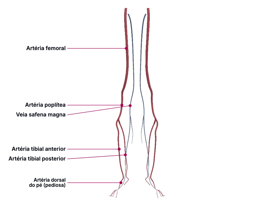
Da virilha ao dorso do pé, os pulsos femoral, poplíteo, tibial posterior e pedioso formam uma cadeia que localiza o nível de uma obstrução arterial na doença arterial periférica - um pulso poplíteo presente com o pedioso ausente aponta uma lesão distal ao joelho, e a claudicação intermitente reproduz esse raciocínio pela distância e pelo músculo comprometido. O índice tornozelo-braquial formaliza essa cadeia num número, comparando a pressão sistólica do tornozelo (tibial posterior ou pedioso) com a do braço. A veia safena magna, por um trajeto subcutâneo longo e previsível do dorso do pé até a virilha, é o principal vaso do sistema venoso superficial - sítio clássico de varizes quando suas próprias valvas falham, e o enxerto mais usado quando uma artéria em outro lugar precisa de bypass.
Educational material written with AI support and checked by automated review, without a named medical reviewer. It does not replace the textbook or clinical guidance.
Essential
A artéria femoral comum nasce como continuação direta da ilíaca externa: 'The common femoral artery arises as a continuation of the external iliac artery after it passes under the inguinal ligament.' Ela fica medial ao ponto médio do ligamento inguinal: 'The common femoral artery is located just medial to the midpoint of the inguinal ligament in the inguinal crease region.' e passa bem em cima da cabeça do fêmur, o que a torna superficial e palpável nesse ponto: 'The average common femoral artery measures approximately 4 cm in length and lies just anterior to the femoral head'
É justamente essa artéria - na sua porção superficial, distal à profunda femoral - o local mais comum de placa aterosclerótica de todo o membro inferior: 'In individuals with peripheral arterial disease (PAD) the most common sites of involvement are the superficial femoral artery and the common femoral artery.' e, de outra fonte que descreve o mesmo achado sobre claudicação: 'The most common location of plaque formation in peripheral arterial disease is in the superficial femoral artery, which may lead to IC symptoms in the calf.' Isso faz do pulso femoral o PRIMEIRO ponto de checagem da cadeia: se ele já está diminuído ou ausente, a obstrução é proximal a ele (aortoilíaca) ou nele mesmo - se está presente e forte, a doença (se houver) está mais distal.
A artéria poplítea é a continuação direta da femoral superficial depois do hiato do adutor, correndo profundamente pela fossa poplítea: 'The popliteal artery is a continuation of the superficial femoral artery as it passes through the adductor hiatus of the adductor magnus muscle, traveling posteriorly to the knee and anterior to its accompanying vein, the popliteal vein, until it bifurcates into the anterior tibial artery and the common trunk of the posterior tibial and peroneal arteries.'
Por estar mais profunda que a femoral, o pulso poplíteo exige uma manobra diferente para ser encontrado: 'the popliteal pulse is palpable in the popliteal fossa with the knee in moderate flexion' E é exatamente essa posição - entre o femoral (proximal) e os tibiais/pedioso (distais) - que dá ao pulso poplíteo seu papel diagnóstico: 'It is important for the evaluation of perfusion to the lower leg in the event the dorsalis pedis and posterior tibial arteries are non-palpable, such as in a person with advanced peripheral artery disease.'
Perto do fim da fossa poplítea, a artéria poplítea se divide em dois ramos terminais: 'Near the distal extent of the popliteal fossa, the popliteal artery divides into the ATA and the tibioperoneal (tibiofibular) trunk.' Um deles, a tibial anterior, desce pelo compartimento anterior da perna e, ao cruzar o tornozelo, muda de nome: 'The dorsalis pedis artery (DPA) is a main artery of the foot. It is a continuation of the anterior tibial artery as it crosses the ankle joint.'
É por ser superficial nesse ponto que a pediosa é o local preferido para o pulso do pé: 'Because the DPA is superficial and readily palpable, it is a preferred site for palpating the pedal pulse on physical examination.' com uma técnica de palpação específica: 'The pedal pulse is palpated by placing the index and third fingers lateral to the extensor hallucis longus tendon and with light compression of the dorsum of the foot, halfway between the first and second metatarsals.'
O outro ramo terminal da poplítea, o tronco tibiofibular, se bifurca por sua vez: 'The tibioperoneal trunk subsequently bifurcates approximately 2.5 cm distal to its origin into the fibular artery and the PTA.' A tibial posterior é o maior dos dois ramos que saem dali: 'The PTA is the largest terminal branch of the popliteal artery' e fica superficial de novo perto do tornozelo: 'At the level of the ankle, the PTA assumes a more superficial course and is palpable posterior to the medial malleolus as the vessel passes through the tarsal tunnel.'
Essa acessibilidade atrás do maléolo medial é o que sustenta seu uso no exame físico: 'The PTA becomes superficial and is readily palpable posterior to the medial malleolus. This anatomical accessibility supports its use in physical examination for assessment of peripheral vascular disease, which affects approximately 12% of the population.' Junto com a pediosa, a tibial posterior é uma das DUAS artérias do tornozelo usadas para calcular o índice tornozelo-braquial: 'The ABI is measured using 3 arteries: the brachial artery for the upper extremity and the dorsalis pedis and or posterior tibial artery at the ankle.' - o ITB formaliza em número a mesma lógica de localização que os dedos do examinador fazem ao palpar.
A veia safena magna é a maior veia do corpo humano: 'The GSV is the longest vein in the body and a major component of the superficial venous system of the lower extremity.' Ela nasce no próprio dorso do pé: 'The vessel originates in the foot at the confluence of the dorsal vein of the first digit and the dorsal venous arch.' e segue um trajeto longo e PREVISÍVEL: 'From its origin, the GSV courses anterior to the medial malleolus, ascends along the medial aspect of the lower leg adjacent to the saphenous nerve, and crosses the posterior border of the medial femoral condyle at the knee before continuing along the anteromedial thigh.' até terminar na virilha: 'The vein traverses the saphenous opening in the fascia lata at the saphenofemoral junction and ultimately drains into the femoral vein within the femoral triangle'
Junto com a pequena safena, ela forma todo o sistema venoso SUPERFICIAL do membro inferior - diferente do sistema arterial dos outros vasos deste painel: 'The superficial venous system consists of the great saphenous vein (GSV) and small saphenous vein, in addition to several accessory veins.' Como qualquer veia de membro inferior, depende de valvas unidirecionais para o sangue subir contra a gravidade: 'Throughout the deep and superficial veins are a number of one-way bicuspid valves that allow blood to travel toward the heart while preventing it from returning to the feet.' É justamente nessa veia - no segmento infrapatelar - que essas valvas mais comumente falham: 'The most common site for the incompetent venous valve is the below-knee great saphenous vein.'
In the clinic
Clinicamente, isso aparece como um padrão de dor: 'Symptoms that indicate femoral artery disease may include claudication (pain or cramping sensation) in the thighs and calves' A localização da dor confirma o nível: 'Patients with aortoiliac disease often experience pain in the buttocks or thighs, while those with femoropopliteal disease most commonly report calf pain.' e 'disease below the inguinal ligament commonly produces claudication affecting the calf muscles' - ou seja, a MESMA lógica de 'onde dói' que localiza a obstrução por pulso também localiza pela topografia da claudicação.
Na prática, isso monta uma régua de dois degraus: femoral presente + poplíteo ausente localiza a obstrução no PRÓPRIO segmento femoropoplíteo; femoral presente + poplíteo presente + pedioso/tibial posterior ausentes localiza a obstrução mais distal, no nível tibial. O exame físico da claudicação formaliza essa checagem sequencial: 'The physical examination should include an assessment of femoral, popliteal, dorsalis pedis, and posterior tibial artery pulses.'
Um pulso pedioso ausente pode indicar doença, mas nem sempre: 'A diminished or absent dorsalis pedis artery pulse may show vascular insufficiency; however, due to differences in the anterior tibialis termination, some individuals may have a congenitally non-palpable pulse.' - por isso a fonte de claudicação intermitente recomenda cautela na interpretação isolada: 'the presence of strong pedal pulses argues against a diagnosis of intermittent claudication' (ou seja, pedioso forte torna a claudicação MENOS provável, mas pedioso fraco isolado não fecha o diagnóstico sozinho - precisa da tibial posterior e do ITB junto).
Um pulso tibial posterior diminuído ou ausente já é, por si só, um sinal de alerta: 'A diminished or absent PTA pulse may indicate vascular insufficiency.' e o ITB traduz isso numericamente: 'Normal: 0.9 to 1.4' é a faixa normal, enquanto 'Low: less than 0.9 means narrowing of vessels'
Quando as valvas falham, o resultado é a varize clássica: 'Varicose veins are characterized by subcutaneous dilated, tortuous veins greater than or equal to three millimeters, involving the saphenous veins, saphenous tributaries, or non-saphenous superficial leg veins' e, se a hipertensão venosa persiste, a pele sofre: 'Persistent venous hypertension can result in hyperpigmentation of the skin, lipodermatosclerosis of the subcutaneous tissue, and, eventually, ulceration.' O tratamento cirúrgico da própria veia doente é a excisão na junção com a femoral: 'The excision of GSV with high ligation at the saphenofemoral junction is sustainable and applicable to all CEAP classes with GSV reflux.' Mas o MESMO trajeto longo e previsível que a torna propensa a essas valvas incompetentes é o que faz dela o enxerto preferido quando uma ARTÉRIA (não ela mesma) precisa de bypass: 'One of the most common surgical interventions is bypass surgery, which reroutes blood flow around a blocked artery using either an autologous vein graft (typically the great saphenous vein) or a synthetic conduit.' - a mesma veia, mas um SEGMENTO SAUDÁVEL dela (não o trecho doente removido na excisão), colhido cirurgicamente e usado como material de reparo na outra situação.
Open the deeper layer
Going deeper
O eixo arterial primitivo do broto do membro inferior, que nasce, junto do plexo terminal, da artéria umbilical: 'The earliest arterial supply to the lower limb bud consists of the axis artery and the terminal plexus, both of which arise from the umbilical artery.' É desse mesmo eixo que saem os ramos que formam duas das artérias desta unidade: 'The axis artery subsequently gives rise to branches that form the ATA and the PTA.'
Artéria tibial anterior - um ramo terminal direto do eixo arterial primitivo, que mais tarde continua como a artéria dorsal do pé (pediosa) depois de cruzar o tornozelo.; Artéria tibial posterior - o outro ramo terminal do mesmo eixo primitivo; já no adulto, dá origem às artérias plantares medial e lateral: 'The ATA continues distally as the dorsalis pedis artery, whereas the PTA gives rise to the medial and lateral plantar arteries.'; Porção proximal da artéria poplítea - um dos segmentos do eixo primitivo que PERSISTEM depois que o restante do vaso embrionário regride: 'Persistent segments contribute to the formation of the inferior gluteal artery, sciatic artery, proximal portion of the popliteal artery, and distal portion of the fibular artery.'; Artéria dorsal do pé (pediosa) - não é, ela mesma, um ramo à parte do eixo primitivo: é a CONTINUAÇÃO da tibial anterior depois que esta cruza o tornozelo. Por isso os pulsos tibial anterior (no músculo tibial anterior, mais acima) e pedioso (no dorso do pé) não são dois vasos diferentes que só coincidem em estar perto um do outro - são o MESMO vaso, com o mesmo eixo primitivo por trás dos dois.; Contraste que NÃO é co-derivado: a artéria femoral nasce SEPARADAMENTE, a partir da ilíaca externa, e só se conecta ao eixo primitivo mais tarde no desenvolvimento: 'A second arterial channel arises independently from the external iliac artery and forms the femoral artery of the lower limb, which subsequently establishes continuity with the axis artery.' A artéria isquiática - o primeiro vaso dominante do embrião, de uma origem totalmente diferente (a umbilical) - regride nesse mesmo período: 'At the 6-mm embryologic stage, the umbilical artery gives rise to the sciatic artery which becomes the dominant blood supply to the developing lower limb.' e 'the iliofemoral system begins to form as the sciatic artery regresses during the 6-33 mm stage' É por isso que um pulso femoral normal, por si só, NÃO garante que a cadeia tibial/pediosa esteja patente: as duas linhagens só se uniram secundariamente, e uma pode estar doente sem que a outra esteja.
Test yourself before moving on
Um paciente com claudicação de panturrilha tem pulso femoral palpável dos dois lados, pulso poplíteo palpável à direita, mas pulso pedioso e tibial posterior ausentes à direita. Em que segmento arterial está a obstrução provável, e por quê?
The answer stays in Assessment, with no spoiler here.
Answer in AssessmentSources (10)
- Swift H, Bordoni B. Anatomy, Bony Pelvis and Lower Limb: Femoral Artery. [Updated 2023 Nov 29]. In: StatPearls [Internet]. Treasure Island (FL): StatPearls Publishing; 2026 Jan-.
The common femoral artery arises as a continuation of the external iliac artery after it passes under the inguinal ligament.
- Patel SK, Surowiec SM. Intermittent Claudication. [Updated 2023 Jul 10]. In: StatPearls [Internet]. Treasure Island (FL): StatPearls Publishing; 2026 Jan-.
The most common location of plaque formation in peripheral arterial disease is in the superficial femoral artery, which may lead to IC symptoms in the calf.
- Zemaitis MR, Boll JM, Kato M, et al. Peripheral Arterial Disease. [Updated 2026 Jan 31]. In: StatPearls [Internet]. Treasure Island (FL): StatPearls Publishing; 2026 Jan-.
This condition results from progressive arterial narrowing, most often in the aortoiliac, femoropopliteal, and infrapopliteal segments, leading to reduced or impaired blood flow to the lower extremities.
- Bowers Z, Nassereddin A, Sinkler MA, et al. Anatomy, Bony Pelvis and Lower Limb: Popliteal Artery. [Updated 2023 Jul 24]. In: StatPearls [Internet]. Treasure Island (FL): StatPearls Publishing; 2026 Jan-.
The popliteal artery is a continuation of the superficial femoral artery as it passes through the adductor hiatus of the adductor magnus muscle, traveling posteriorly to the knee and anterior to its accompanying vein, the popliteal vein, until it bifurcates into the anterior tibial artery and the co…
- Robinson SA, Carlin R. Anatomy, Bony Pelvis and Lower Limb: Foot Dorsalis Pedis Artery. [Updated 2023 Sep 4]. In: StatPearls [Internet]. Treasure Island (FL): StatPearls Publishing; 2026 Jan-.
The dorsalis pedis artery (DPA) is a main artery of the foot. It is a continuation of the anterior tibial artery as it crosses the ankle joint.
- Lezak B, Launico MV. Anatomy, Bony Pelvis and Lower Limb: Posterior Tibial Artery. [Updated 2026 May 24]. In: StatPearls [Internet]. Treasure Island (FL): StatPearls Publishing; 2026 Jan-.
The tibioperoneal trunk subsequently bifurcates approximately 2.5 cm distal to its origin into the fibular artery and the PTA.
- McClary KN, Massey P. Ankle Brachial Index. [Updated 2023 Jan 16]. In: StatPearls [Internet]. Treasure Island (FL): StatPearls Publishing; 2026 Jan-.
The ABI is measured using 3 arteries: the brachial artery for the upper extremity and the dorsalis pedis and or posterior tibial artery at the ankle.
- McMahon K, Anand S, Loyd JW. Saphenous Vein Cutdown. [Updated 2026 Aug 16]. In: StatPearls [Internet]. Treasure Island (FL): StatPearls Publishing; 2026 Jan-.
The GSV is the longest vein in the body and a major component of the superficial venous system of the lower extremity.
- Singh A, Zahra F. Chronic Venous Insufficiency. [Updated 2023 Apr 27]. In: StatPearls [Internet]. Treasure Island (FL): StatPearls Publishing; 2026 Jan-.
The superficial venous system consists of the great saphenous vein (GSV) and small saphenous vein, in addition to several accessory veins.
- Antani MR, Dattilo JB. Varicose Veins. [Updated 2023 Aug 8]. In: StatPearls [Internet]. Treasure Island (FL): StatPearls Publishing; 2026 Jan-.
The most common site for the incompetent venous valve is the below-knee great saphenous vein.
Where this shows up in practice
1 clinical case in the library requires this topic. See where it decides the management.
Ferida crônica na UBS: a úlcera de perna que não é venosa, a mancha que ninguém testou, e a que mudou de aspecto
Medicina de família e comunidade
Open the case for free
More Anatomia summaries
After reading, use what you learned
Atlas
See the structures of the topic in 3D and move through the layers.
Clinical case
Apply the content to an on-call decision.
Exam
Read the result that closes the reasoning.
Assessment
Answer the block questions and see where you stand.
AI tutor
Ask about what the text did not settle.
Start free: create an account and try a sample of the 3D Atlas, the clinical case, the exam, the assessment and the AI tutor. No credit card.
Start free